기흉
Pneumothorax
■ 기흉의 원인
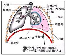
늑막 강 속에 공기가 차 있는 상태를 기흉이라 한다.
갓 태어난 신생아([부모도 반의사가 되어야 한다–소아가정간호백과]-제 6권 신생아 성장 발육 양호 질병–기흉 참조)와 신생아기 이후 아이에게 생기는 기흉의 원인이 다를 수 있다(그림 112 기흉 참조).
확실한 원인 없이 생기는 1차성 자연 기흉(Primary spontaneous pneumothorax)과 이미 존재한 질병과 관련되어 생긴 2차성 기흉(Secondary spontaneous pneumothorax)으로 분류 한다.
1차성 자연 기흉은 남성 만 명 중 7.4, 여성 만 명 중 1.2명 발생한다.
체질적으로 외배엽 체형을 가진 사람들에게 좀 더 생길 수 있다.
1차성 자연 기흉이 있었던 가족 병력이 있는 사람들에게 더 잘 생긴다(참고문헌:Contemporary pediatrics, December, 2008. p.504).
황색 포도상구균성 폐렴(p.00 폐렴 참조)·기관지 천식·폐결핵, 만성 차단성 폐질환, 에이즈, 뉴모 시스티스 폐렴, 그 밖에 다른 여러 종류의 폐렴·폐농양·폐낭포·폐기흉·흉곽수술·흉곽 외상 등으로 2차성 기흉이 생길 수 있다.
다른 병이 있고 그 병으로 인해 생긴 기흉을 2차성 기흉이라고 한다.
아무런 이유도 없이 기흉이 생길 수 있다. 이런 기흉을 1차성(원발성) 자연 기흉이라고 한다.
참고로, 늑막 강 내 압은 정상 적으로 음압이고, 기관지 내 압이나 폐기포 내 압은 정상 적으로 양압이다.
기흉이 있는 늑막강 내 압이 기관지 내 압이나 폐기포 내 압보다 더 높을 때가 있다.
기흉 압이 비정상적으로 상당히 높고 기흉이 상당히 클 때는 기흉이 있는 쪽의 폐가 기흉으로 눌려 쪼그라질 수 있다. 이런 기흉을 긴장성 기흉(Tension pneumothorax)라 한다(그림 113 참조).
■ 기흉의 증상 징후
기흉의 종류와 중증도, 원인, 합병증 유무 등에 따라 증상 징후가 다르다.
흡인 폐렴(p.00 신생아 흡인성 폐렴 참조), 황색 포도상구균성 폐렴, 기관지 천식 등으로 생긴 2차성 기흉의 증상 징후는 원래 있던 병으로 생긴 증상 징후와 2차성 기흉으로 생긴 증상 징후와 함께 나타난다.
긴장성 기흉이 있을 때는 갑자기 호흡곤란이 생길 수 있고, 안색이 창백하면서 안절부절 못하고 기흉이 생긴 쪽 가슴이 아플 수 있다.
기흉이 경미할 때는 아무런 증상 징후가 없을 수 있다.
1차성 자연 기흉이 있을 때는 기흉이 있는 쪽 가슴이 갑자기 아프고 숨이 가쁘고 빈맥이 생기고 숨소리가 감소되고 기침을 할 수 있다.
■ 기흉의 진단
병력·증상 징후·진찰소견 등을 종합해서 이 병이 의심되면, 가슴 X-선 사진 검사로 진단할 수 있다.
기흉을 일으킨 원인에 따라 진단 한다.
■ 기흉의 치료
기흉의 종류, 중증도, 기흉을 일으킨 원인에 따라 치료한다.
1차성 자연 기흉의 일부는 약 1~2주일 동안 육체적 안정을 취하면 자연히 치료될 수 있다.
주사바늘 등으로 늑막강 내 있는 공기를 빼주고 관찰하면 자연적으로 회복될 수도 있다.
심한 긴장성 기흉은 생명에 위험할 수 있다. 늑막 강 속 공기를 흉벽 관으로 빼주고 쪼그라진 폐를 정상적으로 회복시키기는 치료도 한다.
때로는 작은 흉벽 관이나 큰 주사 바늘을 기흉이 있는 늑막강 속에 넣고 기흉 공기를 응급으로 빼는 치료도 한다.
기흉이 있는 늑막 강 속에 넣었던 흉벽 관은 기흉 공기가 더 이상 나오지 않을 때까지 계속 삽입 해 놓았다가 기흉 공기가 더 이상 나오지 않으면 빼는 식으로 치료하기도 한다.
이런 치료 방법으로 완치가 안 되면 늑막 강 속으로 공기가 들어가게 하는 폐 환부나 세기관지 환부를 적절히 외과적으로 치료 한다.
기흉을 일으킨 원인을 동시 치료한다.
전문적인 면도 있지만 소아청소년 자녀 양육에 많은 도움이 되리라고 믿습니다. 그러나 여기에 있는 정보는 여러분의 의사로부터 얻는 정보 진단 치료를 대신할 수 없습니다.
부모도 반의사가 되어야 한다
Pneumothorax
Causes of pneumothorax
A condition in which air is filled in the pleural cavity is called pneumothorax.
The causes of pneumothorax that occur in a newborn newborn (see [Parents should also be half-doctors – Pediatric and Home Nursing Encyclopedia] – Volume 6 Diseases for Newborn Growth and Development – Pneumothorax) and those that occur in children after the neonatal period may be different (see Figure 112 Pneumothorax). .
It is classified into primary spontaneous pneumothorax, which occurs without a clear cause, and secondary spontaneous pneumothorax, which occurs due to an existing disease.
Primary spontaneous pneumothorax occurs in 7.4 out of 10,000 men and 1.2 out of 10,000 women.
It is more likely to occur in people with a constitutionally ectomorphic body type.
It is more likely to occur in people with a family history of primary spontaneous pneumothorax (Reference: Contemporary pediatrics, December, 2008, p.504).
Staphylococcus aureus pneumonia (see p. 00 pneumonia), bronchial asthma, pulmonary tuberculosis, chronic obstructive pulmonary disease, AIDS, pneumocystis pneumonia, and various other types of pneumonia, lung abscess, lung cyst, pneumothorax, and thoracic surgery. Secondary pneumothorax may occur due to thoracic trauma.
A pneumothorax caused by another disease is called a secondary pneumothorax.
Pneumothorax can occur for no reason. This kind of pneumothorax is called primary (primary) spontaneous pneumothorax.
For reference, intrapleural pressure is normally negative, and intrabronchial or alveolar pressure is normally positive.
There are times when the pressure within the pleural space with pneumothorax is higher than the pressure within the bronchi or alveoli.
When the pneumothorax pressure is abnormally high, and the pneumothorax is quite large, the lung on the side where the pneumothorax is located may be compressed and shrunk by the pneumothorax. This type of pneumothorax is called tension pneumothorax (see Figure 113).
■ Symptoms and signs of pneumothorax
Symptoms and signs vary depending on the type, severity, cause, and presence of pneumothorax complications.
Symptoms of secondary pneumothorax caused by aspiration pneumonia (see p. 00 Neonatal aspiration pneumonia), Staphylococcus aureus pneumonia, bronchial asthma, etc. appear together with the symptoms caused by the original disease and the symptoms caused by secondary pneumothorax.
When you have a tension pneumothorax, you may suddenly have difficulty breathing, your complexion may become pale and restless, and the chest on the side where the pneumothorax occurred may be painful.
When pneumothorax is mild, there may be no symptoms.
When there is a primary spontaneous pneumothorax, the chest on the pneumothorax side may suddenly become painful, short of breath, tachycardia may occur, breath sounds may decrease, and coughing may occur.
■ Diagnosis of pneumothorax
If this disease is suspected based on the history, symptoms, signs, and examination findings, the diagnosis can be made with a chest X-ray.
Diagnosis is made according to the cause of the pneumothorax.
■ Treatment of pneumothorax
Treatment is based on the type, severity, and cause of the pneumothorax.
Some cases of primary spontaneous pneumothorax can be cured naturally by taking physical rest for about 1 to 2 weeks.
Natural recovery may occur if the air in the pleural cavity is removed with a needle and observed.
Severe tension pneumothorax can be life-threatening. Treatment is also used to remove air from the pleural cavity through chest wall tubes and restore shrunken lungs to normal.
Sometimes, a small chest wall tube or a large needle is inserted into the pleural space where the pneumothorax is located and the pneumothorax air is removed as an emergency treatment.
The chest wall tube inserted into the pleural cavity where the pneumothorax is located may be treated by continuing to insert it until the pneumothorax air no longer comes out, and then removing it when the pneumothorax air no longer comes out.
If complete cure is not possible with these treatment methods, the affected lung or bronchioles that allow air to enter the pleural cavity should be treated surgically appropriately.
Treat the cause of pneumothorax simultaneously.
Although it has a professional aspect, I believe it will be of great help in raising children and adolescents. However, the information here is not a substitute for information, diagnosis, and treatment from your doctor.
www.drleepediatrics.com
Parents should also become half-doctors (Encyclopedia of Pediatric and Family Nursing)
Copyright drleepediatrics, com 2/l7/.2026