늑막염(흉막염)과 농흉 Pleurisy and empyema
For more information, please visit drleepediatrics.com. and then Search.
정보를 더 찾으려면 drleepediatrics.com 방문한 후 Search 에서 찾아보세요.
■ 늑막염(흉막염)과 농흉의 개요 및 원인
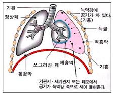
폐는 두 겹의 엷은 막으로 둘러싸여 있다.
이 두 겹의 엷은 막을 늑막 또는 흉막이라고 한다.
폐 표면을 둘러싼 흉막을 폐흉막 또는 폐늑막이라 하고, 흉곽의 내면에 접촉돼있고 폐늑막의 바깥 면에 접해 있는 늑막을 벽흉막이라고 한다.
이 두 겹의 늑막(흉막) 사이에 있는 공간을 늑막 강(흉막 강)이라고 한다(그림 111참조).
늑막 강 속에는 미끈거리는 체액이 정상적으로 소량 들어있다.
결핵균이나 다른 종류의 박테리아, 또는 그 외 다른 병원체에 늑막 강이 1차적으로 감염되면 늑막강 속에 감염병이 생길 수 있다.
이렇게 생긴 늑막 강 감염병을 늑막염(Pleurisy/흉막염)이라고 한다.
늑막 강에 박테리아 감염이 생기고 그로 인해 늑막 강 속에 감염병이 생기면 늑막 강 감염균의 종류에 따라 짙은 혼탁 고름, 엷은 혼탁 고름, 또는 맑은 혼탁 고름이 늑막 강 속에 괼 수 있다.
또 병원체가 감염되지 않았는데 신증후군이나 심장병 등으로 전신의 각 계통의 각 기관과 조직에 체액이 비정상적으로 괼 수 있다.
이런 경우, 늑막 강 속에 맑은 체액이 괼 수 있다. 즉 늑막 강 속, 복강 속, 그 외 신체의 다른 계통의 다른 조직과 기관에 체액이 비정상적으로 많이 괼 수 있다.
화농성 세균이 늑막 강에 감염되고 그로 인해 늑막 강 감염병이 생기면 늑막 강 속에 고름이 잡힐 수 있다. 이때 늑막 강 속에 짙고 혼탁한 고름이 괸 늑막염이 생길 수 있다.
이런 늑막염을 농흉(Empyema)이라고 한다.
폐렴을 앓는 소아청소년 150명 중 1명에게 농흉이 생길 수 있고 소아 청소년 입원 환아 1000명 증 0.4~6명에게 농흉이 있었다.
최근에는 소아 청소년들에게 농흉이 더 자주 발견되고, 특히 폐렴연쇄상구균 폐렴이 소아 청소년들에게 있을 때 가슴 초음파 검에 8~14%에서 농흉이 발견된다고 한다(출처: Pediatric News, March 2004).
박테리아, 바이러스, 그 밖의 다른 병원체가 늑막염 및, 또는 농흉을 일으킬 수 있다.
황색 포도상구균,
A군 베타 용혈성 연쇄상구균,
B형 헤모필러스 인플루엔자균,
폐렴연쇄상구균,
그 밖에 다른 종류의 박테리아가 늑막 강에 직접 감염되거나 간접 감염되어 늑막염(흉막염)이나 농흉을 일으킬 수 있다.
폐렴을 일으킨 박테리아가 늑막 강 속으로 1차적으로 감염되어 늑막염이나 농흉을 일으킬 수 있다.
결핵균, 바이러스, 마이코플라스마 등 병원체가 직접 또는 간접적으로 늑막강 속이나 폐에 일차적으로 감염되어 늑막염을 일으킬 수 있다.
그런 늑막염으로 생긴 늑막 강 속 체액은 맑고 투명한 것이 보통이다.
췌장염, 요독증, 신증후군, 심장병, 간경변증, 악성 종양, 백혈병 등을 앓을 때도 능막강 속에 맑고 투명한 체액이 2차적으로 괼 수 있다.
■ 늑막염(흉막염)과 농흉의 증상 징후
늑막염이나 농흉을 일으킨 원인, 다른 동반 병의 유무, 합병증의 유무, 병의 중증도에 따라 증상 징후가 다르다.
박테리아 감염으로 늑막염이 생겼을 때는 늑막염이 생긴 쪽의 가슴이 아프고 열이 나며 호흡곤란이 생긴다.
늑막염과 폐렴이 동시에 생길 수 있다. 이때 두 가지 병으로 생기는 증상 징후가 함께 나타날 수 있다.
■ 늑막염(흉막염)과 농흉의 진단
증상 징후, 진찰소견, 가슴 X-선 사진, 피 검사와 소변 검사, 세균 배양검사 등을 종합하여 진단할 수 있다.
늑막염이 결핵균 감염으로 생겼을 때는 결핵 피부 반응검사 결과가 양성으로 나타난다.
원인을 더 확실하게 알아보기 위해 늑막 강 내 체액이나 고름을 빼고 그 고름으로 그람 염색 현미경 세균 검사, 세균 배양검사 및 다른 여러 가지 검사로 진단할 수 있다.
■ 늑막염(흉막염)과 농흉의 치료
늑막염을 일으킨 원인과 합병증의 유무 등에 따라 치료한다.
결핵균에 의한 늑막염이 있을 때는 결핵 치료약으로 치료한다.
농흉의 경과를 3기로 나눌 수 있다. 각기에 따라 치료를 달리할 수 있다.
황색 포도상구균 감염이나 다른 종류의 박테리아 감염으로 생긴 농흉이 있을 때는 늑막 강에 있는 고름을 흉막 튜브(관)를 통한 배농 치료 하거나
흉막천자 수술 배농 치료,
섬유소 융해제 치료, 외과적 배농 방법으로 고름을 빼 주는 동시 적절한 항생제 정맥주사로 치료할 수 있다.
신증후군, 심장병 등으로 늑막 강 속에 늑막 액이 괼 때는 신증후군이나 심장병 등 원발성 원인이 되는 병을 치료하면 늑막 강 속에 괴었던 체액이 자연히 빠져 없어진다.
늑막강 내 Urokinase 주입 치료 또는 비디오를 통한 흉막강 내시경 외과적 처치로 치료 할 수 있다.
출처: Pediatric News
부모도 반의사가 되어야 한다
Pleurisy and Empyema
For more information, please visit drleepediatrics.com and then search.
■ Overview and Causes of Pleurisy and Empyema
The lungs are surrounded by two layers of thin membranes.
These two layers of thin membranes are called the pleura.
The pleura surrounding the surface of the lung is called the visceral pleura, and the pleura in contact with the inner surface of the chest cavity and adjacent to the outer surface of the visceral pleura is called the parietal pleura.
The space between these two layers of pleura is called the pleural cavity (see Figure 111).
Normally, a small amount of slippery fluid is present in the pleural cavity.
If the pleural cavity is primarily infected with tuberculosis bacteria, other types of bacteria, or other pathogens, an infectious disease can develop in the pleural cavity.
This infectious disease of the pleural cavity is called pleurisy.
If a bacterial infection occurs in the pleural cavity, and an infectious disease develops in the pleural cavity as a result, depending on the type of bacteria infecting the pleural cavity, thick purulent fluid, thin purulent fluid, or clear purulent fluid may accumulate in the pleural cavity.
Also, even without a pathogen infection, fluid can abnormally accumulate in various organs and tissues of the body due to conditions such as nephrotic syndrome or heart disease.
In such cases, clear fluid may accumulate in the pleural cavity. That is, an abnormally large amount of fluid may accumulate in the pleural cavity, abdominal cavity, and other tissues and organs of other systems of the body. When pyogenic bacteria infect the pleural cavity, leading to a pleural cavity infection, pus can accumulate in the pleural space. This can result in pleurisy with a thick, cloudy accumulation of pus in the pleural cavity.
This type of pleurisy is called empyema.
Empyema can occur in 1 in 150 children and adolescents with pneumonia, and 0.4 to 6 out of 1000 hospitalized children and adolescents had empyema.
Recently, empyema has been found more frequently in children and adolescents, and it is reported that empyema is detected in 8-14% of cases of pneumococcal pneumonia in children and adolescents during chest ultrasound examinations (Source: Pediatric News, March 2004).
Bacteria, viruses, and other pathogens can cause pleurisy and/or empyema.
Staphylococcus aureus,
Group A beta-hemolytic streptococcus,
Haemophilus influenzae type B,
Streptococcus pneumoniae,
and other types of bacteria can directly or indirectly infect the pleural cavity, causing pleurisy or empyema.
Bacteria that cause pneumonia can primarily infect the pleural cavity, leading to pleurisy or empyema.
Tuberculosis bacteria, viruses, mycoplasma, and other pathogens can directly or indirectly infect the pleural cavity or lungs, primarily causing pleurisy.
The fluid in the pleural cavity resulting from such pleurisy is usually clear and transparent. Clear, transparent fluid can also accumulate secondarily in the pleural cavity in cases of pancreatitis, uremia, nephrotic syndrome, heart disease, cirrhosis, malignant tumors, and leukemia.
■ Symptoms and Signs of Pleurisy (Pleural Effusion) and Empyema
The symptoms and signs vary depending on the cause of pleurisy or empyema, the presence of other co-existing diseases, the presence of complications, and the severity of the disease.
When pleurisy is caused by bacterial infection, there is pain in the chest on the affected side, fever, and shortness of breath.
Pleurisy and pneumonia can occur simultaneously. In this case, the symptoms and signs of both diseases may appear together.
■ Diagnosis of Pleurisy (Pleural Effusion) and Empyema
Diagnosis can be made by comprehensively considering symptoms and signs, physical examination findings, chest X-ray images, blood and urine tests, and bacterial culture tests.
When pleurisy is caused by tuberculosis infection, the tuberculin skin test result will be positive.
To determine the cause more definitively, fluid or pus can be extracted from the pleural cavity, and the pus can be used for Gram staining microscopy, bacterial culture tests, and various other tests for diagnosis.
■ Treatment of Pleurisy (Pleural Effusion) and Empyema
Treatment depends on the cause of pleurisy and the presence of complications.
When pleurisy is caused by tuberculosis bacteria, it is treated with tuberculosis medication.
The course of empyema can be divided into three stages. Treatment may vary depending on each stage. When empyema is caused by Staphylococcus aureus infection or other types of bacterial infections, treatment involves draining the pus from the pleural cavity through a pleural tube (catheter),
or through thoracentesis,
fibrinolytic therapy, or surgical drainage, along with appropriate intravenous antibiotic treatment.
When pleural effusion occurs in the pleural cavity due to nephrotic syndrome or heart disease, treating the underlying primary disease, such as nephrotic syndrome or heart disease, will naturally resolve the accumulated fluid in the pleural cavity.
Treatment can also involve intrapleural urokinase injection or video-assisted thoracoscopic surgery.
Source: Pediatric News
Parents should also become half-doctors.
Copyright drleepediatrics.com 2/17/2026
|