척주 측만증(척추 옆굽음증)
Scoliosis
■ 척주 측만증의 원인
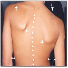
▴ 사진 129. 척주 측만. 점선으로 표시된 바와 같이 척주가 측만되어 있다. 옆으로 쭉 팔과 허리 사이의 간격에 차이가 난다(a, b 사이). 양쪽 어깨 높이가 다르다.
Copyrightⓒ 2001 John Sangwon Lee, MD.FAAP
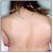
▴ 사진 130. 척주 측만이 현저하게 되어 있다. 사춘기 여아의 후면 사진.
Copyrightⓒ 2001 John Sangwon Lee, MD.FAAP
● 척주 만곡에는 경추 만곡, 흉추 만곡, 요추 만곡, 골반 만곡이 정상적으로 있다.
● 척주의 일부가 비정상적으로 좌측 옆으로나 우측 옆으로 만곡 된 상태를 척주 측만이라 한다.척주 측만으로 생기는 증상을 척주 측만증(Scoliosis) 또는 척주 측만곡증이라 한다.
● 학령기 아이들에게 척주 측만증이 생기는 빈도는 거의 10%이다.
● 척주 측만증 중 약 60~80%는 특발성 척주 측만증이다.
● 척주 측만증에는 선천성으로 생기는 선천성 척주 측만증과 후천성으로 생기는 후천성 척주 측만증이 있다.
● 원인을 확실히 알 수 없는 원발성 척주 측만증(특발성 척주 측만증),
● 근육신경 이상으로 생기는 근육신경 이상 척주 측만증 등이 있다.
● 평소에 자세를 바르게 하지 않아 생기는 척주 측만증, 척주염이나 척주 외상이나 척주에 생긴 종양 등으로 척주가 비정상으로 좌측 옆으로나 우측 옆으로 만곡 되어 생기는 경도 척주 측만증도 있다.
● 원발성 척주 측만증은 생후 어느 때든지 생길 수 있다. 그러나 사춘기가 시작되기 바로 전에 더 잘 생긴다.
● 여아의 경우 11세경, 남아의 경우 12~14세경에 척주 측만증의 증상 징후가 현저하게 나타나는 것이 보통이다.
● 즉 사춘기가 시작되기 전부터 조금 만곡 되어 있던 척주 측만이 사춘기가 오기 시작하면 척주가 1~2년 동안에 더 현저히 더 심하게 만곡 되어 척주가 더 뚜렷하게 측만 될 수 있고 증상 징후도 더 현저히 나타 날 수 있다.
● 뇌성마비, 소아마비, 척수 수막류, 프리드라히 운동실조, 척수외상, 신경섬유증, 마르팡증, 엘러스단로스증, 척수종양, 척주골종, 구루병, 불완전 골형성증, 고 비타민 A증, 갑상선 기능저하증, 연소성 류마토이드 관절염, 뮤코다당체 침착증(점다당질증), 척주염, 척주 결핵 등으로 척주 측만증이 생길 수 있다.
■ 척주 측만증(척추 측만곡증)의 증상 징후
● 척주 측만증을 일으킨 원인과 정도에 따라 증상 징후가 다르다.
● 이론적으로 척주의 어느 부위에도 생길 수 있다.
● 흉부 척주에만 생길 수도 있고 흉부 척주의 일부와 허리 척주(요부 척주)의 일부에만 생길 수도 있고, 어떤 때는 허리 척주(요부 척주)에만 생길 수 있다.
● 척주 측만이 생긴 척주 부위가 좌측으로 또는 우측으로 경미하게 만곡될 수 있고, 심하게 만곡될 수 있다.
● 척주 측만의 정도가 심하지 않을 때는 척주가 조금 측만되었을 뿐 아무런 다른 증상 징후가 나타나지 않는다.
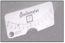
▴ 사진 132. 척주 측만곡의 정도를 재는 척추 측만 측정 기구(척추 측만계).
Copyright ⓒ 2011 John Sangwon Lee, MD. FAAP\
● 척주 측만이 심하게 생겼을 때는 외관상으로 척주 측만이 있는 것을 쉽게 알 수 있다. 사진 129~130 참조.
● 더 심하면 옷을 입고 있는 상태에서도 척주가 측만된 것이 겉으로 나타나고 평소에 운동을 자유자재로 할 수 없다.
● 심한 척주 측만을 적절히 치료해 주지 않으면 흉강 구조에도 변화가 생겨서 폐 기능 장애도 생기고 그로 인해 호흡 곤란이 생길 수 있고 수명이 더 짧아질수 있다. 소스:NEJM Journal Watch 5/2015
■ 척주 측만증의 진단
● 척주 측만증의 주증도에 따라 경증, 중등증, 중증의 척주 측만증으로 구분한다.
● 병력, 증상 징후와 진찰소견 등을 종합해 척주가 측만되어 있다고 의심되면 척추측만계로 척주 측만의 정도를 재보고 필요에 따라 척주 X선 사진 검사로 확진한다.
● 경증 척주 측만이 있는 유아들, 학령기 아이들, 사춘기 아이들이 옷을 입고 있을 때는 이 병이 있는지 쉽게 알 수 없다.
● 겉옷을 벗고 반듯이 서 있을 때 머리끝에서 엉덩이까지 척주 전체를 살펴보면 척주의 일부가 우측 옆으로 또는 좌측 옆으로 휘어지고
● 한쪽 어깨의 높이가 다른 쪽 어깨 높이보다 더 낮다.
● 한쪽 앞가슴이 다른 쪽 앞가슴보다 앞으로 더 불쑥 나와 있다.
● 반듯이 서서 두 팔을 양쪽 옆구리로 반듯이 내려 양 옆 몸통에 붙일 때 한쪽 옆구리와 그쪽 팔 사이의 간격이 반대쪽의 사이 간격보다 더 많이 벌어져서 양쪽 옆구리와 팔 사이의 간격이 다르다(사진 129 참조).
● 척추측만계로 척주 만곡의 정도를 쟀을 때 척주가 6~7도 이상 만곡되면 척주 만곡이 확실히 있는 것으로 진단한다.
● 마지막으로 척주 X선 사진 검사로 척주 측만의 정도를 더 확실히 알아보고,
● 또 육체적 운동, 물리치료, 브레이스, 수술로 치료해야 하는지 알아본다.
■ 척주 측만증의 치료
● 정도에 따라 다르게 치료한다.
● 경증 척주 측만이 있을 때는 자신도 부모도 자녀가 그 병을 가졌는지 잘 알지 못할 때가 많다. 경증 척주 측만이 불과 몇 달 동안에 중증 척주 측만으로 변화할 수 있다.
● 소아청소년 정기 건강검진을 받을 때마다 척주 측만이 있는지 기본적으로 진찰해서 알아야 한다.
● 경증 척주 측만을 조기에 진단해서 더 이상 악화되지 않도록 조기에 적절히 치료해야 한다.
● 경증 척주 측만은 적절한 운동과 물리치료 등으로 치료한다.
● 경증 척주 측만을 처음으로 진단받은 후부터 적어도 만 18세가 될 때까지 더 악화되는지 주기적으로 추적 진찰을 받고 필요에 따라 적절한 치료도 받아야 한다.
● 경증 척주 측만이 짧은 기간 동안에 더 악화되어 중등도 척주 측만이나 중증 척주 측만으로 될 수 있다.
● 그때그때 척주 측만의 진행 정도에 따라 운동, 물리치료, 찰스턴(Charleston) 브레이스 등으로 적기에 적절히 치료받아야 한다. 이렇게 해서 될 수 있는 한 수술 치료를 피해야 한다.
● 중증 척주 측만은 밀워키 브레이스 또는 척수 수술 등으로 치료한다.
● 브레이스 치료를 해 줄 때는 목 부위에서부터 엉덩이 부위까지 걸치는 브레이스로 2~4년간 밤낮으로 치료하든지, 또는 찰스턴(Charleston) 브레이스로 낮에만 치료할 수 있다.
● 선천성 척주 측만은 3~4세에 수술로 치료할 수 있고,
● 근육신경성 척주 측만증은 브레이스 치료로 잘 치료되지 않고 수술로 치료하는 것이 일반적이다. 오랫동안 이런 치료를 받을 때 육체적으로 정신적으로 큰 부담이 될 수 있다.
● 그러므로 척주 측만은 조기에 진단하여 가능한 한 운동치료나 물리치료 등으로 보다 더 쉽고 덜 고생하면서 치료받아야 한다.
● 즉 브레이스 치료나 척수 수술로 치료받아야 할 때까지 척주 측만증이 진행되지 않도록 최대한 노력해야 한다.
● 중기나 후기의 사춘기 아이들에게 생기는 특발성 척주 측만증의 예후는 일반적으로 좋다.
● 척추 측만증의 증도에 따라, 척주 측만증을 치료할 때 쓰는 브레이스로 인해 호흡곤란도 생길 수 있다.
● 이런 측만 척주증을 치료할 때 쓰는 브레이스로 호흡기 기능에 많은 제한이 생길 수 있다.
Copyright drleepediatrics.com 2/27/2026
Scoliosis (Curved Spine)
■ Causes of Scoliosis
▴ Photo 129. Scoliosis. The spine is curved, as indicated by the dotted line. There is a difference in the distance between the arms and the waist (a and b). The shoulder heights are different on both sides.
Copyrightⓒ 2001 John Sangwon Lee, MD.FAAP
▴ Photo 130. Prominent scoliosis. Posterior view of an adolescent girl.
Copyrightⓒ 2001 John Sangwon Lee, MD.FAAP
● Normal spinal curvatures include cervical, thoracic, lumbar, and pelvic curvatures.
● Scoliosis is an abnormal curvature of the spine, either to the left or right. The symptoms caused by scoliosis are called scoliosis or scoliosis.
● Scoliosis occurs in approximately 10% of school-age children.
● Approximately 60-80% of scoliosis cases are idiopathic.
● Scoliosis can be either congenital or acquired.
● Primary scoliosis (idiopathic scoliosis) is caused by unknown causes,
● Myoneural scoliosis is caused by neuromuscular disorders.
● Mild scoliosis is caused by poor posture, and spondylitis, spinal trauma, or tumors in the spinal column, causing an abnormal curvature of the spine to the left or right.
● Primary scoliosis can occur at any time after birth, but it is more common just before puberty.
● In girls, the symptoms of scoliosis typically become noticeable around age 11, and in boys, around ages 12 to 14.
● In other words, a slightly curvature of the spine before puberty can become more pronounced over the course of 1 to 2 years as puberty begins. This can lead to a more pronounced scoliosis and more pronounced symptoms.
● Scoliosis can be caused by cerebral palsy, poliomyelitis, myelomeningocele, Friedreich’s ataxia, spinal cord trauma, neurofibromatosis, Marfan’s disease, Ehlers-Danlos disease, spinal tumors, spinal osteoma, rickets, osteogenesis imperfecta, hypervitaminosis A, hypothyroidism, juvenile rheumatoid arthritis, mucopolysaccharidosis (spondylosis), spondylitis, and spinal tuberculosis.
■ Symptoms and Signs of Scoliosis
● Symptoms and signs of scoliosis vary depending on the cause and severity.
● Theoretically, scoliosis can occur anywhere in the spine.
● It can occur only in the thoracic spine, or in part of the thoracic spine and part of the lumbar spine, or sometimes only in the lumbar spine.
● The affected spine can be slightly curved to the left or right, or it can be severe.
● When scoliosis is mild, the spine is only slightly curvatured and no other symptoms or signs appear.
▴ Photo 132. A scoliometer, a device used to measure the degree of scoliosis.
Copyright ⓒ 2011 John Sangwon Lee, MD. FAAP
● When scoliosis is severe, it is easily recognizable visually. See photos 129-130.
● In more severe cases, scoliosis is visible even while wearing clothing, hindering normal movement.
● If severe scoliosis is not properly treated, changes in the thoracic structure can occur, leading to lung dysfunction, respiratory distress, and a shortened lifespan. Source: NEJM Journal Watch 5/2015
■ Diagnosis of Scoliosis
● Depending on the severity of the scoliosis, it is classified as mild, moderate, or severe.
● If scoliosis is suspected based on medical history, symptoms, signs, and physical examination findings, the degree of scoliosis is measured with a scoliometer and, if necessary, confirmed with a spinal X-ray.
● Mild scoliosis is not easily detectable in infants, school-aged children, or adolescents while wearing clothing. ● When standing upright with your outer clothing removed, examining the entire spine from head to hips reveals a portion of the spine curved to the right or left.
● One shoulder is lower than the other.
● One chest protrudes further forward than the other.
● When standing upright and placing both arms straight down to your sides and touching them to your sides, the distance between one side of your body and that arm is greater than the distance between the other side, resulting in a difference in the distance between the arms and your sides (see Photo 129).
● When measuring the degree of the spinal curvature with a scoliometer, a spinal curvature of 6-7 degrees or more is diagnosed as definitive.
● Finally, a spinal X-ray examination is performed to further determine the degree of scoliosis.
● We also determine whether physical exercise, physical therapy, bracing, or surgery are necessary for treatment.
■ Treatment of Scoliosis
● Treatment varies depending on the severity. ● When children have mild scoliosis, neither they nor their parents are often aware that they have it. Mild scoliosis can progress to severe scoliosis in just a few months.
● Children should be screened for scoliosis at every regular pediatric and adolescent health checkup.
● Mild scoliosis should be diagnosed early and treated appropriately to prevent further worsening.
● Mild scoliosis is treated with appropriate exercise and physical therapy.
● From the initial diagnosis of mild scoliosis, children should receive regular follow-up examinations to monitor for further worsening until at least age 18, and receive appropriate treatment as needed.
● Mild scoliosis can worsen over a short period of time, developing into moderate or severe scoliosis.
● Depending on the severity of scoliosis, appropriate treatment should be provided in a timely manner, including exercise, physical therapy, and a Charleston brace. This approach can help avoid surgery whenever possible. Severe scoliosis is treated with a Milwaukee brace or spinal surgery.
Brace treatment can be performed day and night for 2-4 years with a brace that extends from the neck to the hips, or with a Charleston brace, which can be performed only during the day.
Congenital scoliosis can be treated surgically at ages 3-4.
Muscular-neurogenic scoliosis is less effective with braces and is typically treated surgically. Long-term treatment can be physically and mentally taxing.
Therefore, scoliosis should be diagnosed early and treated with exercise therapy or physical therapy whenever possible, making treatment easier and less painful.
In other words, every effort should be made to prevent scoliosis from progressing until it requires brace therapy or spinal surgery.
The prognosis for idiopathic scoliosis, which occurs in children during mid- to late-puberty, is generally good. Depending on the severity of scoliosis, braces used to treat scoliosis can also cause respiratory distress.
Braces used to treat scoliosis can significantly limit respiratory function.
Copyright drleepediatrics.com 2/27/2026