발목 관절뼈 골절과 발뼈 골절 Ankle joint bone fractures and Foot bone fracture
- 운동, 장난, 또는 안전사고 등으로 발목 관절(족관절) 뼈나 발뼈 등의 일부 또는 전부가 골절될 수 있다.
- 발목 관절(Ankle joint)은 경골 최 하부, 비골 최 하부, 거골, 종골 등 족근골, 연골, 활막, 인대, 근육, 건, 혈관 등으로 구성되어 있다.
- 발목 관절에 있는 뼈의 일부 또는 전부가 골절 될 수 있고,
- 발목 관절을 구성하고 있는 연골이나 인대, 또는 건 등이 손상되어 발목이 삘 수 있고 탈구될 수 있다.
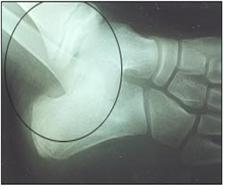
사진 171. ◯내 심하게 탈골된 발목 관절 및 골절된 발목 관절
Copyright ⓒ 2011 John Sangwon Lee, M.D.. FAAP
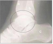
사진 172. 정상 발목 관절과 발목 관절에 있는 족근골 측면 X-선 사진
a-경골, b-비골, c-종골, ◯내–발목 관절
Copyright ⓒ 2011 John Sangwon Lee, M.D., FAAP
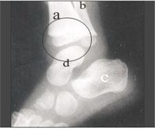
사진 173. 정상 발목 관절과 족근골의 측면 X-선 사진
a-경골, b-비골, c-종골(발꿈치뼈), d-거골(목말 뼈),e-입방골, f-주상골 ◯내–발목 관절
Copyright ⓒ 2011 John Sangwon Lee, M.D., FAAP
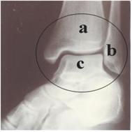
사진 174. ◯내–정상 발목 관절 X-선 사진
a-경골,
b-비골,
c-종골,
d-목말 뼈(거골)
Copyright ⓒ 2011 John Sangwon Lee, M.D., FAAP
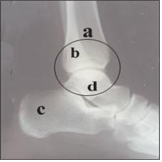
사진 175. ◯내–정상 발목 관절 측면 X-선 사진
a-경골, b-비골, c-종골, d-목말 뼈, e-입방골, f-주상골
Copyright ⓒ 2013 John Sangwon Lee, M.D., FAAP
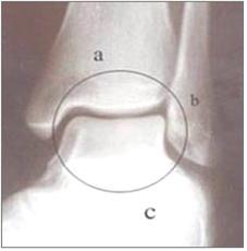
사진 176. 정상 발목 관절 X-선 사진 전 면
a-경골, b-비골, c-종골, ◯내–발목 관절
Copyright ⓒ 2011 John Sangwon Lee, M.D., FAAP
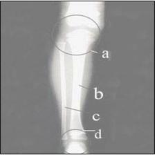
사진 177. 정상 발목 관절과 경골과 비골의 전면 X-사진
a ◯내–무릎 관절, b-경골, c-비골, d-발목 관절
Copyright ⓒ 2011 John Sangwon Lee, M.D., FAAP
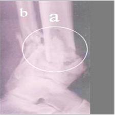
사진 178. 경골, 비골, 발목 관절뼈에 생긴 심한 복합 골절
a-경골, b-비골, ◯내–발목 관절
Copyright ⓒ 2011 John Sangwon Lee, M.D., FAAP
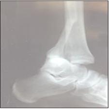
사진 179. 비골과 발목 관절뼈의 골절의 측면 X선 사진
a-경골, b-비골, c-종골, ◯내–비골 골절
Copyright ⓒ 2011 John Sangwon Lee, M.D., FAAP
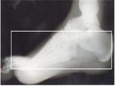
사진 180. 발뼈의 측면 X-선 사진
□내 뼈들은 발뼈.
발뼈=족근골+중족골+족지골
Copyright ⓒ 2011 John Sangwon Lee, M.D., FAAP
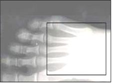
사진181. 중족골과 발가락뼈(족지골)의 전후면 X- 선 사진
□내 뼈들은 족지골의 일부와 중족골의 전두면 사진
Copyright ⓒ 2011 John Sangwon Lee, M.D., FAAP
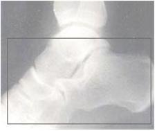
사진182. □내 뼈들은 발 족근골
Copyright ⓒ 2011 John Sangwon Lee, M.D., FAAP
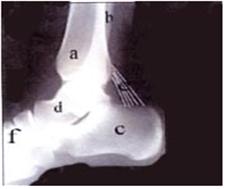
사진183. 하각(아랫다리), 발목 관절뼈와 족근골 측면 x선 사진
a-경골, b-비골, c-종골, d-목말뼈(거골), e-아킬레스 건, f-중족골
Copyright ⓒ 2011 John Sangwon Lee, M.D., FAAP
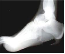
사진 184. 발뼈(족근골+중족골+족지골)의 전후면 X-선 사진
Copyright ⓒ 2011 John Sangwon Lee, M.D., FAAP
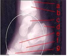
사진 185. ◯내– 발뼈 측면 X-선 사진
a-비골, b-경골, c-거골(목말뼈, 발목뼈), d-종골(발꿈치뼈), e-주상골, f-설상골, g-중족골
Copyright ⓒ 2011 John Sangwon Lee, M.D., FAAP
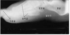
사진 186. 발뼈와 발바닥의 측면 x선 사진
Copyright ⓒ 2011 John Sangwon Lee, M.D., FAAP
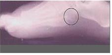
사진 187. ◯내 발뼈가 골절됐다.
Copyright ⓒ 2011 John Sangwon Lee, M.D., FAAP
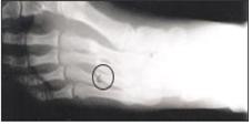
사진 188. ◯내 넷째 중족골이 골절됐다.
Copyright ⓒ 2011 John Sangwon Lee, M.D., FAAP
- 발목 관절뼈가 골절될 때 발목 관절 내에 있는 한 개의 뼈만 골절 될 수도 있지만 발목 관절을 구성하고 있는 여러 종류의 뼈들 중 한 개나 그 이상 여러 개의 뼈가 동시 골절될 수 있다.
-
발목 관절을 형성하고 있는 인대, 활막, 연골, 근육, 건 등이 손상될 수 있다.
-
발목 관절이 삘 때 외과(외측 복사뼈)가 제일 흔히 골절된다.
-
여기서는 발뼈 골절이나, 발목뼈 골절에 관해 주로 설명한다.
발목 관절뼈나 발뼈 골절의 증상 징후
-
발목 관절뼈나 족근골에 생긴 골절의 정도, 그 중 어느 뼈에 골절이 생겼는지에 따라, 발목 관절을 이루고 있는 연골, 인대, 건, 활막, 혈관, 말초 신경 등의 손상 정도에 따라 증상 징후가 다르다.
-
발목 관절뼈나 족근골 골절로 생기는 증상 징후는 발목이나 발이 삐었을 때나 발목 속에 있는 뼈가 탈골될 때 생길 수 있는 증상 징후와 거의 비슷할 수 있다.
-
일반적으로 골절된 뼈가 있는 발목 관절 부위나 발의 부위가 붓고 아프고 그쪽 발목이나 발 기능의 장애가 생길 수 있다.
-
그쪽 발목이나 발을 능동적으로 움직이기 싫어한다.
-
정상적으로 움직일 수 있는 최대한도 범위로 움직이지 못한다.
-
수동적으로 그쪽 발목이나 발을 움직일 때 아파한다.
-
골절이 있는 발목 관절 부위를 손으로 누르면 아프고 자신이나 다른 사람이 골절된 족근골이 있는 발의 부위나 골절된 발목 관절뼈가 있는 발목뼈 부위로 움직이면 그 부위가 더 아플 수 있다.
-
발목 관절뼈나 족근골이 골절 됐을 때 골절되어 있는 발로 정상적으로 서고 걷지 못한다. 그쪽 발에 힘주고 서기를 싫어한다.
-
골절된 후 2~3일정도 지나면 골절된 부위가 더 심하게 붓고 멍들 수 있다.
-
국소에 따뜻한 촉감이 있지만 전신 열은 나지 않는 것이 보통이다.
발목 관절뼈나 발뼈 골절의 진단
-
병력, 증상 징후와 진찰소견 등을 종합해 발목 관절뼈나 족근골이 골절되었다고 의심되면 발 x-선 사진 검사나 발목 X-선 사진 검사로 진단한다.
-
발 뼈나 발목 관절 속 뼈가 골절되었을 때 발이나 발목이 타박상 등 단순히 외상만 입었는지, 발목 관절 속이 삐었는지, 또는 발목 관절 속에 있는 뼈가 탈구되어 있는지 그냥 눈으로 보고 진찰해 감별 진단을 할 수 없는 때도 많다.
-
즉 발목 관절 속 뼈만 또는 족근골만 골절 되었는지, 발목이 삐었는지, 발목 관절 좌상이 생겼는지를 감별 진단해야 한다.
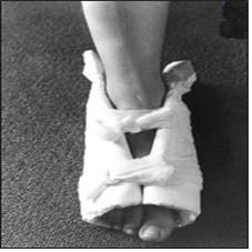
사진 189. 족근골, 중족골 또는 족지골 등 발뼈에 골절이 생겼다고 의심되면 두꺼운 패드나 베개 등을 발에 대어 발을 고정시킬 수 있다.
Copyright ⓒ 2011 John Sangwon Lee, M.D., FAAP
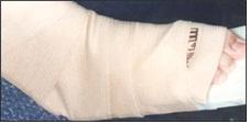
사진 190. 발목 관절뼈나 발뼈 골절이 생겼다고 의심하면 두꺼운 패드나 베개 등으로 발과 발목을 고정시킬 수 있다.
Copyright ⓒ 2011 John Sangwon Lee, M.D., FAAP
발목 관절뼈나 발뼈 골절의 치료
-
발목 관절뼈가 골절되었다고 의심되면 그쪽 발이나 발목관절로 더 이상 걷지 않게 하고 환아를 안정시킨다.
-
동시 의료구급대원이나 병원 응급실에 긴급 전화 진료상담을 하고 그들의 지시에 따라 현장에서 치료를 시작한다.
-
의료구급대원이나 의사가 현장으로 와 응급치료를 할 수 없는 경우에는 큰 베개나 스펀지 등으로 관절뼈 골절이 있다고 의심하는 쪽 발, 발목과 아랫다리 전체를 적절히 받치고 골절된 발이나 발목이 더 이상 움직이지 못하게 부목으로 고정시킨 후 구급차로나 다른 적절한 교통수단을 이용해 적절한 병원 응급실이나 근처 병원으로 이송한다.
-
골절된 발목이나 발을 당기거나 밀거나 비틀지도 말고 가능한 한 골절된 당시 처음 목격한 상태 그대로 계속 유지하면서 골절된 발목이나 발에 부목을 댄다.
-
때 의사에게 긴급 전화 진료 상담을 해서 그의
-
지시에 따라 응급처치를 하는 것이 이상적이다.
-
발이나 발목 관절뼈가 골절되었을 때 가능하면 발이나 아랫다리의 혈액순환이 정상인지 신경 기능에 이상이 있는지 체크해보고 필요에 따라 의사에게 보고하고 그의 지시대로 응급 처치한다.
-
병원 응급실로 이송 중 발목 관절뼈가 골절되어 있는 쪽의 발톱 밑 혈색이 정상인지 발등 동맥 맥박이 정상적으로 뛰는지 체크해서 발과 발목의 혈액순환이 정상인지 알아보고 필요에 따라 의사에게 전화 보고한다.
-
발등 동맥 맥박이 정상적으로 뛰지 않거나 그 쪽의 발톱 밑 혈색이 파랗거나 창백할 때는 의사나 의료구급대원이나 병원 응급실에 긴급 전화를 해 그 사실을 보고하고 그들의 지시에 따라 구급차 등을 이용해 응급실로 더 급히 이송한다.
Ankle joint bone fractures and Foot bone fracture 발목 관절뼈 골절과 발뼈 골절
• Some or all of the ankle joint (ankle joint) bones or foot bones may be fractured due to exercise, play, or safety accidents.
• The ankle joint is composed of the lowermost part of the tibia, the lowermost part of the fibula, the talus, the calcaneus, etc.
• Some or all of the bones in the ankle joint may be fractured;
• The cartilage, ligaments, or tendons that make up the ankle joint may be damaged and the ankle may be sprained or dislocated.
Picture 171. ◯The severely dislocated ankle joint and fractured ankle joint Copyright ⓒ 2011 John Sangwon Lee, M.D., FAAP
Picture 172. Lateral X-ray of the tarsal bone in the normal ankle joint and the ankle joint a-tibia, b-fibula, c-calcaneus, ◯ inner-ankle joint Copyright ⓒ 2011 John Sangwon Lee, M.D., FAAP
Picture 173. Lateral X-ray of normal ankle joint and tarsal bone a-tibia, b-fibula, c-calcaneus (heel bone), d-talus (talus bone), e-cubic bone, f-navicular bone ◯inner-ankle joint Copyright ⓒ 2011 John Sangwon Lee, M.D., FAAP
Picture 174. ◯Inner-normal ankle joint X-ray a – tibia, b – fibula, c – calcaneus, d – talus bone (talus) Copyright ⓒ 2011 John Sangwon Lee, M.D., FAAP
Picture 175. ◯The intra-normal ankle joint lateral X-ray a – tibia, b – fibula, c – calcaneus, d – the tibia, e – cuboid, f – navicular Copyright ⓒ 2013 John Sangwon Lee, M.D., FAAP
Picture 176. Anterior view of the normal ankle joint X-ray a-tibia, b-fibula, c-calcaneus, ◯ inner-ankle joint Copyright ⓒ 2011 John Sangwon Lee, M.D., FAAP
Picture 177. Anterior X-photograph of the normal ankle joint and tibia and fibula an ◯ Intra-knee joint, b-tibia, c-fibula, d-ankle joint Copyright ⓒ 2011 John Sangwon Lee, M.D., FAAP
Picture 178. Severe complex fractures in the tibia, fibula, and ankle articular bones a-tibia, b-fibula, ◯in-ankle joint Copyright ⓒ 2011 John Sangwon Lee, M.D., FAAP
Picture 179. Lateral X-ray of fracture of fibula and ankle articular bone a-tibia, b-fibula, c-calcaneus, ◯in-fibular fractures Copyright ⓒ 2011 John Sangwon Lee, M.D., FAAP
Picture 180. Lateral X-ray of the foot bone □My bones are my feet. Foot bones = tarsal bones + metatarsal bones + foot bones Copyright ⓒ 2011 John Sangwon Lee, M.D., FAAP
Picture 181. Anterior and posterior X-ray of metatarsal and toe bones (phalanges) □My bones are part of the phalanges and frontal photos of the metatarsals Copyright ⓒ 2011 John Sangwon Lee, M.D., FAAP
Picture 182. My bones are the tarsal bones of the feet Copyright ⓒ 2011 John Sangwon Lee, M.D., FAAP
Picture 183. X-ray image of the lower leg (lower leg), ankle joint and tarsal bones a – tibia, b – fibula, c – calcaneus, d – talus (talus), e – Achilles tendon, f – metatarsal Copyright ⓒ 2011 John Sangwon Lee, M.D., FAAP
Picture 184. Anterior and posterior X-ray of foot bones (tarsal bones + metatarsal bones + foot phalanges) Copyright ⓒ 2011 John Sangwon Lee, M.D., FAAP
Picture 185. ◯Inner-lateral X-ray of the foot bone a-fibula, b-tibia, c-talus (neck, ankle), d-calcaneus (heel), e-navicular, f- cuneiform, g-metatarsal Copyright ⓒ 2011 John Sangwon Lee, M.D., FAAP
Picture 186. Lateral x-ray of the foot bones and soles Copyright ⓒ 2011 John Sangwon Lee, M.D., FAAP
Picture 187. ◯My foot bone was fractured. Copyright ⓒ 2011 John Sangwon Lee, M.D., FAAP
Picture 188. ◯My fourth metatarsal bone was fractured. Copyright ⓒ 2011 John Sangwon Lee, M.D., FAAP
• When an ankle joint bone is fractured, only one bone within the ankle joint may be fractured, but one or more bones among various types of bones constituting the ankle joint may be fractured simultaneously.
• The ligaments, synovial membrane, cartilage, muscle, and tendon that form the ankle joint may be damaged.
• Surgical (lateral malleolus) fractures are the most common fractures of the ankle joint.
• This section mainly discusses ankle fractures and ankle fractures.
Symptoms, signs of an ankle joint or foot fracture
• Symptoms differ depending on the degree of fracture in the ankle joint or tarsal bone, which bone is fractured, and the degree of damage to the cartilage, ligaments, tendons, synovial membrane, blood vessels, and peripheral nerves that make up the ankle joint.
• The symptoms of an ankle joint or tarsal fracture can be very similar to those that can occur when an ankle or foot is sprained or a bone in the ankle is dislocated.
• In general, the area of the ankle joint or foot with fractured bones is swollen and painful, and there may be impairment of ankle or foot function.
• Dislikes actively moving that ankle or foot.
• Inability to move within the maximum range of normal movement.
• Pain when passively moving the ankle or foot.
• It hurts when you press on the fractured ankle joint, and it can be more painful if you or someone else moves to the area of the foot with the fractured tarsal bone or the area of the ankle with the fractured ankle joint.
• When the ankle joint or tarsal bone is fractured, you cannot stand and walk normally with the fractured foot. He doesn’t like to stand with his feet strained.
• Two or three days after the fracture, the fractured area may become more swollen and bruised.
• There is usually a feeling of warmth in the area, but nobody heat.
Diagnosis of an ankle joint or foot bone fracture
• If it is suspected that the ankle joint or tarsal bone is fractured based on the medical history, symptom signs, and examination findings, a foot X-ray examination or an ankle X-ray examination is performed.
• When a bone in the foot or ankle joint is fractured, a differential diagnosis can be made by simply looking and examining whether the foot or ankle is simply traumatized, such as a bruise, a sprained ankle joint, or a bone in the ankle joint is dislocated. Many times there is no
• In other words, it is necessary to differentially diagnose whether only the bones in the ankle joint or only the tarsal bones are fractured, whether the ankle is sprained, or whether the ankle joint has sprained.
Photo 189. If you suspect that a fracture has occurred in the foot bones, such as the tarsal bone, metatarsal bone, or phalanx, you can immobilize the foot by placing a thick pad or pillow against the foot. Copyright ⓒ 2011 John Sangwon Lee, M.D., FAAP
Photo 190. If you suspect that you have fractured the ankle joint or foot bone, you can immobilize the foot and ankle with a thick pad or pillow. Copyright ⓒ 2011 John Sangwon Lee, M.D., FAAP
Treatment of fractures of the ankle joint or foot bones
• If it is suspected that the ankle joint is fractured, stop walking with that foot or ankle joint and stabilize the child.
• Simultaneously provide emergency phone consultations to paramedics or hospital emergency departments and initiate on-site treatment according to their instructions.
• If medical paramedics or doctors cannot come to the scene and provide emergency treatment, use a large pillow or sponge to properly support the foot, ankle, and the entire lower leg on the side of the suspected joint bone fracture, and the fractured foot or ankle will no longer heal. Immobilize them with a splint and transport them to an appropriate hospital emergency room or nearby hospital by ambulance or other suitable means of transportation.
• Splint the fractured ankle or foot while avoiding pulling, pushing, or twisting the fractured ankle or foot and, if possible, keep it as it was first seen at the time of the fracture. • When to call your doctor for an emergency phone consultation
• It is ideal to provide first aid as directed.
• When a foot or ankle joint bone is fractured, if possible, check whether the blood circulation in the foot or lower leg is normal or if there is an abnormality in nerve function, and if necessary, report it to the doctor and provide first aid according to his instructions.
• During transport to the hospital emergency room, check whether the blood under the toenail on the side where the ankle joint bone is fractured is normal or the pulse of the dorsal artery is beating normally, check whether the blood circulation in the foot and ankle is normal, and call the doctor if necessary.
• When the pulse of the dorsal artery is not beating normally, or the color under the toenail on that side is blue or pale, make an emergency call to a doctor, paramedic, or hospital emergency room to report the fact, and use an ambulance, etc.
transport
출처 및 참조문헌
- www.drleepediatrics.com 제16권 소아청소년 정형외과 질환
- www.drleepediatrics.com제8권 소아청소년 호흡기 질환
- www.drleepediatrics.com제9권 소아청소년 소화기 질환
- www.drleepediatrics.com제10권. 소아청소년 신장 비뇨 생식기 질환
- www.drleepediatrics.com제11권. 소아청소년 심장 혈관계 질환
- www.drleepediatrics.com제12권. 소아청소년 신경 정신 질환, 행동 수면 문제
- www.drleepediatrics.com제13권. 소아청소년 혈액, 림프, 종양 질환
- www.drleepediatrics.com제14권. 소아청소년 내분비, 유전, 염색체, 대사, 희귀병
- www.drleepediatrics.com제15권. 소아청소년 알레르기, 자가 면역질환
- Red book 29th-31st edition 2021
- Nelson Text Book of Pediatrics 19th — 21st Edition
- The Johns Hopkins Hospital, The Harriet Lane Handbook, 22nd edition
-
Quick Reference to Pediatric Emergencies, Delmer J. Pascoe, M.D., p.80-81, 164-165
-
Emergency Pediatrics, A guide to ambulatory care, 5th edi. Roger M. Barkin, Peter Rosen, p.552-555
-
Emergency care and transportation of the sick and injured, 3rd edition, American Academy of orthopedic surgeons. p.134, 163-164, 162-163
-
Nelson textbook, 19th edition
-
Childhood Emergencies in the Office, Hospital and Community, American Academy of Pediatrics
-
Emergency Medical Service for Children, By Ross Lab. May 1989. p.10
-
Emergency care, Harvey grant and Robert Murray
-
Emergency Care Transportation of Sick and Injured American Academy of Orthopaedic Surgeons
-
Emergency Pediatrics A Guide to Ambulatory Care, Roger M. Barkin, Peter Rosen
-
Immediate care of the acutely ill and injured, Hugh E. Stephenson, Jr
-
The Critically Ill Child, Diagnosis and Management, Edited by Clement A. Smith
-
Emergency Medical Services for Children: The Role of the Primary Care Provider, America Academy of Pediatrics
-
Quick Reference To Pediatric Emergencies, Delmer J. Pascoe, M.D., Moses Grossman, M.D. with 26 contributors
-
Manual of Emergency Care
-
응급환자관리 정담미디어
-
소아가정간호백과–부모도 반의사가 되어야 한다, 이상원
-
Neonatal Resuscitation American heart Association
-
Neonatology Jeffrey J.Pomerance, C. Joan Richardson
-
Pediatric Resuscitation Pediatric Clinics of North America, Stephen M. Schexnayder, M.D.
-
Pediatric Critical Care, Pediatric Clinics of North America, James P. Orlowski, M.D.
-
Preparation for Birth. Beverly Savage and Dianna Smith
- Infectious disease of children, Saul Krugman, Samuel L Katz, Ann A. Gerhon, Catherine Wilfert
-
The Harriet Lane Handbook 19th Edition
-
소아과학 대한교과서
-
제1권 소아청소년 응급의료 참조문헌과 출처
-
Other
Copyright ⓒ 2015 John Sangwon Lee, MD., FAAP
“부모도 반의사가 되어야 한다”-내용은 여러분들의 의사로부터 얻은 정보와 진료를 대신할 수 없습니다.
“The information contained in this publication should not be used as a substitute for the medical care and advice of your doctor. There may be variations in treatment that your doctor may recommend based on individual facts and circumstances. “Parental education is the best medicine.”