www.drleepediatrics.com 제1권 소아청소년 응급의료 제 2부 제16장 피부 발진과 열이 나는 질병들(1) Diseases with skin rash and fever(1)
- 소아청소년들(0~18세)에게 열도 나고 피부에발진이 생기는 병들이 많다.
- 여기서는 소아청소년들에게 열도 나고 피부에 발진이 나는 많은 병들 중 일부를 소개한다.
- 소아청소년들에게서 비교적 흔히 볼 수 있는 피부 발진의 형태와 종류, 원인에 관해 피부 발진 사진을 통해 이미 설명했다.
- 피부에 발진만 나고 열이 나지 않는 질환과
- 열은 나지만 피부에 발진이 나지 않는 질환,
- 피부 발진도 나고 열이 나는 질환에 관해 다음 각 항에서 요약해서 설명한다.
피부 발진의 여러 종류의 형태 (사진 2 참조)
|
|
소아청소년들(0~18세)에게 피부 발진만 나든지, 또는 열이 나고 피부 발진도 나면 그 피부 발진은 다음 1.~29.에 해당되는 감염병이나 그 외 질병에 속할 가능성이 있다. 드물게는 그 다음 여러 가지 감염병이나 질병들 중 한 종류, 두 종류, 또는 그 이상 여러 종류의 질병으로 피부 발진이 생길 수 있다. 물론 여기서 열거한 감병이나 질병 이 외 다른 병으로 피부발진이 생길 수 있다.
|
1. 성홍열 Scarlet Fever
-
A군 베타 용혈성 연쇄상구균 감염으로 생긴 인두염이나 인두편도염이 있을 때 성홍열이 생길 수 있다.
-
드물게는 피부 상처에 생긴 A군 베타 용혈성 연쇄상구균 감염이 있을 때 성홍열이 생길 수 있다.
-
인두염이나 인두편도염을 일으킨 A군 베타 용혈성 연쇄상구균의 균종에서 나오는 균 독소로 인해 성홍열 피부발진이 생긴다.
-
열이 나고 인두통, 팔다리와 전신이 아프고 구토와 두통 등의 증상 징후가 생길 수 있다.
-
홍반 구진 발진이 얼굴, 목, 가슴, 배, 손 등에 생길 수 있다.
-
그 피부 발진은 사포와 같이 도돌도돌할 수 있다. 이런 감염병을 성홍열이라고 한다.
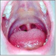
그림 76. A군 베타 용혈성 연쇄상구균성 편도선염과 성홍열.
붉은 딸기 모양의 혀는 성홍열의 증상들 중 하나이다.
Copyright ⓒ 2011 John Sangwon Lee, MD., FAAP
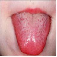
그림 75. 성홍열로 인한 붉은 딸기 모양 혀.
Copyright ⓒ 2011 John Sangwon Lee, MD., FAAP
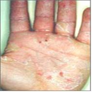
그림 77. 성홍열로 인해 손바닥의 표피가 얇게 벗겨졌다.
Copyright ⓒ 2011 John Sangwon Lee, MD., FAAP
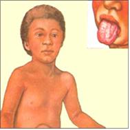
그림 78. 성홍열을 앓을 때 사포와 같이 도돌도돌하고 붉은 피부 발진이 난다.
Health information service, Merk Sharp & Dome, West Point, PA
-
혓바닥의 색이 빨간 딸기 색이나 붉은 소고기 색과 비슷하게 변할 수 있고,
-
인두와 편도가 붉고 편도가 커진다.
-
병력, 증상, 진찰소견 등을 종합해서 이병을 쉽게 진단할 수 있다.
-
A군 베타 용혈성 연쇄상구균 항원 항체 응집반응 검사(Quick Strep Test)나 인두 편도에서 점액을 채취해서 A군 베타 용혈성 연쇄상구균 배양검사로 확진한다.
-
항생제로 치료해야한다.
-
[부모도 반의사가 되어야 한다–소아가정간호백과]-제7권 소아 청소년 감염병–성홍열 참조.
2. 라임 병 및 라임 병성 관절염 Lyme disease and Lyme arthritis
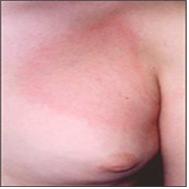
그림 79. 라임 병으로 생기는 만성 유주성 홍반.
Copyright ⓒ 2011 John Sangwon Lee, MD., FAAP
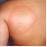
그림 80. 라임 병으로 생기는 만성 유주성 홍반.
Copyright ⓒ 2011 John Sangwon Lee, MD., FAAP
-
볼레리아 불그돌페리(Borrelia Burgdorferi) 박테리아 감염으로 생기는 감염병이다.
-
관절염, 심장염 또는 뇌막염 등이 생길 수 있고 만성 유주성 홍반(Erythema Chronicum Migrans)이 생길 수 있다.
-
만성 유주성 홍반의 모양은 원형, 타원형 등일 수 있다.
-
얼굴, 목, 몸통, 다리 등에 한 개 내지 여러 개가 동시 날 수 있다.
-
그 중앙 부위는 정상 피부색과 같을 수 있다.
-
만성 유주성 홍반의 전체의 색이 붉을 수 있다.
-
라임 병이 있을 때 만성 유주성 홍반도 생기고 라임 병을 일으키는 세균이 피부에 직접 감염되어 1차성 라임 병 피부염이 생길 수 있다. 출처-18
-
병력, 증상 징후, 진찰소견, 라임 병 검사 등으로 진단한다.
-
12세 이전 소아 청소년들의 라임 병은 Amoxicillin 등의 항생제로,
-
12세 이후 소아들의 라임 병은 Doxycyclin 등 항생제로 치료한다.
-
라임 병 참조
3. 수막구균 패혈증 및, 또는 수막구균 뇌막염 Meningococcemia and, or Meningococcal meningitis
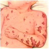
그림 81. 수막구균 패혈증으로 생긴 피부출혈 반점
Used with permission from Ross lab.Columbia Ohio. USA
그림 82. 수막구균 패혈증으로 생긴 자반증과 수포.
출처:10
-
수막구균 패혈증은 수막구균 감염으로 생기는 감염병이다.
-
처음에는 감기의 증상과 비슷하다가 고열, 두통, 저혈압, 혼수, 경련 등의 전신 증상이 심하게 생길 수 있다.
-
이 균이 피부에 직접 감염되어 피부에 수막구균 피부염이 생길 수 있고
-
홍역 발진과 비슷한 홍반성 구진, 점상 출혈반점이 나타날 수 있고,
-
혈소판 감소증과 혈액응고 장애로 전신에 자반증과 점상 출혈반점 등이 생길 수 있다.
-
병력, 증상 징후, 진찰소견, 세균 배양검사 등으로 진단하고 적절한 항생제 혈관주사로 치료한다.
-
p.00 수막구균 패혈증 뇌막염 참고
-
[부모도 반의사가 되어야 한다–소아가정간호백과]-제7권 소아 청소년 감염병–패혈증 참조
4. 전염성 농가진(감염성 농가진) Impetigo contagiosa
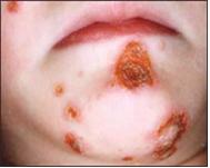
사진 83. 농가진.
Copyright ⓒ 2011 John Sangwon Lee, MD., FAAP
-
A군 베타 용혈성 연쇄상구균 및, 또는 황색 포도상구균 감염으로 생기는 박테리아 피부염이다.
-
얼굴, 목, 사지, 몸통 등의 피부층의 표면에 주로 난다.
-
한 개 또는 여러 개의 농가진이 동시 날 수 있다.
-
홍반, 수포, 농포, 가피(딱지) 등이 생길 수 있다.
-
박트로반(Bactroban) 크림이나 Retapamulin 연고, 또는 Centany(Mupirocin ointment) 2% 연고(출처: Contemporary Pediatrics June 2007) 등으로 농가진 병소가 있는 국부에 발라 치료할 수 있고 Oxacillin 등 경구용 항생제로 치료 할 수 있다.
-
농가진 참조.
5. 포도상 구균 화상 피부 증후군 Staphylococcal scalded skin syndrome(SSSS)
-
황색 포도상구균 외 독소 반응으로 화상피부 증후군이 급성으로 생기는 피부 질환이다.
-
심한 2도 화상을 입은 피부와 같이 피부염이 생긴다.
-
유아들이나 10세 이전 학령기 아이들에게 더 잘 생긴다.
-
홍반성 반점, 표피박리, 탈피, 낙설 등의 병변이 피부에 생기고 권태감, 열, 신경과민 등의 전신 증상이 동시 생길 수 있다.
-
사포 같은 홍반성 발진도 날 수 있다.
-
이런 발진들이 머리카락이 나지 않는 머리부위에서부터 전신으로 다 퍼져 나고 입술 주위와 손가락 사이 등에도 생긴다.
-
병력, 증상 징후, 진찰소견 등을 종합해서 이 병이 의심되면 포도상구균 세균 배양검사 등을 해서 진단한다.
-
Vancomycin, Clindamycin, Oxacillin 등 항 포도상구균 항생제로 치료하고 증상은 대증치료 한다. [부모도 반의사가 되어야 한다–소아가정간호백과]-제8권 소아 청소년 호흡기 질환 참조
6. 홍역 Rubeola(Measles)
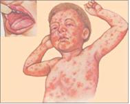
그림 84. 자연 홍역으로 생긴 홍역꽃 피부와 입안 점막에 생긴 코플릭 반점. 출처-Copyrightⓒ 2001 John Sangwon Lee,MD.FAAP
-
홍역바이러스 감염으로 생기는 전신 바이러스 감염병이다.
-
전형적인 홍역의 증상은, 발병 후 첫 24~48 시간 동안 열과 권태감이 계속되다가,
-
기침(Cough),
-
콧물(Coryza),
-
결막염(Conjunctivitis) 의 세 가지 증상(3CS)이 항상 나타난다.
-
-
이 세 가지 증상을 영어로 3Cs 증상이라고 한다.
-
입안 점막에 코플릭크 반점이 난다.
-
그 반점이 나타난 1~2일이 되면 홍반성 구진상 발진(홍역 꽃)이 귓바퀴 뒤 부위에 있는 피부에서부터 나기 시작해서 2~4일 동안에 얼굴, 몸통, 팔 다리 손 등으로 퍼져 난다.
-
그 피부 발진은 5~8일 동안 났던 순서대로 없어지는 것이 보통이다.
-
홍역 예방접종 백신을 접종 받은 후 홍역 면역체가 생성된 후 항체가 체내에서 거의 다 없어진 아이들에게 생긴 경한 홍역, 또는 홍역 예방접종 백신을 받지 않은 아이들에게 자연 홍역 바이러스 감염으로 생기는 자연홍역과 있을 때 홍역 꽃이 서로 다르므로 감별 진단해야 한다.
-
홍역 참조.
-
병력, 증상 징후 , 진찰소견을 종합해서 진단한다. 대증치료 한다.
7. 풍진 Rubella(3 days measles)
-
풍진 바이러스 감염으로 인한 전신 급성바이러스 감염병을 풍진이라 한다.
-
두통,
-
안구가 아프고
-
인두통,
-
미열,
-
한전 등 경미한 증상이 3~4일 동안 계속된다.
-
후두골 아래의 목 부위, 귓바퀴 뒤 부위와 그 아래의 목 부위에 있는 림프절이 비대 되고 아플 수 있다.
-
이 병이 발병한 후 4~5일이면 연분홍색 발진, 홍반성 발진과 구진이 이마와 귀 뒤 바퀴 머리카락이 나지 않는 부위에 나기 시작해서 얼굴, 목, 몸통, 배, 팔다리 등의 피부로 퍼져 난다.
-
이런 반점이 2~3일 경과하면 자연히 없어진다.
-
병력, 증상, 증후, 진찰소견 등을 종합해서 진단한다.
-
임신 첫 3개월 중 이 병에 걸리면 태아가 선천성 풍진 증후군에 걸릴 수 있다.
-
그 태아에게 백내장, 선천성 심장기형, 농아, 폐렴, 간 비대와 비장의 비대, 혈소판 감소증 등 선천성 풍진 증후군이 생길 수 있다.
-
[부모도 반의사가 되어야 한다–소아가정간호백과]-제6권 신생아 성장 발육 양호 질환–선천성 풍진 증후군. 제7권 소아청소년 감염병–풍진 참조
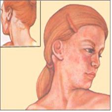
그림 85. 풍진으로 생긴 피부 발진과 귓바퀴 뒤와 두발의 가장자리 부위에 생긴 림프절 비대. 출처; Health information service, Merk Sharp & Dome, West Point, PA
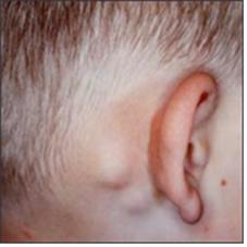
사진 86. 풍진으로 귓바퀴 뒤와 두발 가장자리 부위에 생긴 림프절 비대.
Copyright ⓒ 2011 John Sangwon Lee, MD. FAAP
8. 돌발성 발진(장미진) Exanthem subitum(Roseola)
-
인간 헤르페스 바이러스 6 감염 또는 인간 헤르페스바이러스 7 감염으로 생기는 급성 바이러스 감염병이다.
-
3개월~3세 영유아들에게 주로 생기는 감염병이다.
-
섭씨 39~40도 정도 고열이 1~4일 동안 계속 나다가 그 열이 떨어지자마자 홍역 꽃 비슷하게 또는 풍진 꽃과 비슷한 붉은 홍반 반점, 홍반 구진 발진이 날 수 있다.
-
이 발진이 몸통에서 처음 나기 시작해서 얼굴, 목, 팔다리가 있는 방향으로 퍼져 나는 것이 보통이다.
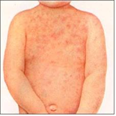
그림 87. 장미진의 피부 발진.
Copyrightⓒ 2001 John Sangwon Lee,MD.FAAP
-
그 발진은 1~2일 동안 진행되다 자연히 없어진다.
-
발진이 나기 전에 고열로 열성경련을 할 수 있다.
-
진찰 상 특이한 증후가 없기 때문에 확진을 하기 곤란할 때가 많다.
-
발병 일에서 열이 떨어질 때까지 상당히 보챌 수 있다.
-
병력, 증상 징후, 진찰소견 등을 종합해서 진단하고 타이레놀로 해열시킨다.
-
[부모도 반의사가 되어야 한다–소아가정간호백과]-제7권 소아 청소년 감염병–돌발성 발진 참조.
9. 전염성 홍반 Erythema Infectiosum(Fifth Disease/Human parvovirus B19 infection and Skin rashes)
-
인간 파보바이러스 B19 감염으로 생기는 바이러스 감염병이다.
-
이병을 제5병 이라고도 한다.
-
열, 두통, 한전, 근육통, 권태감 등의 증상이 경미하게 생길 수 있다.
-
처음에는 양 볼이 붉어지고 그 다음에는 양 어깨와 위팔 등에 홍반성 반점, 구진양 반점이 그물 모양과 비슷하게, 또는 레이스 모양과 같이 날 수 있다.
-
발진이 2~3주 동안 갈 수 있다.
-
관절통이 10% 정도 생긴다.
-
임신부가 이 바이러스에 감염되면 태아에게 심한 빈혈이나 태아 수종증이 생길 수 있다.
-
병력 증상 징후, 진찰소견 등을 종합해서 진단한다.
-
대증 치료를 한다
-
부모도 반의사가 되어야 한다–소아가정간 호백과]-제7권 소아 청소년 감염병 참조.
10. 수두 Chickenpox(Varicella)
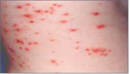
그림91. 수두 피부 발진.
Copyrightⓒ 2001 John Sangwon Lee,MD.FAAP
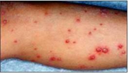
그림90. 수두 피부 발진.
Copyrightⓒ 2001 John Sangwon Lee,MD.FAAP
-
수두바이러스 감염으로 생기는 바이러스 감염병이다.
-
10~20일 간의 잠복기를 거쳐 이 병이 발병된다.
-
미열 내지 고열, 두통 등의 전신 증상이 생길 수 있다.
-
맨 처음에는 몸통에 홍반성 반진이 생기고 그 발진이 구진상 반진으로 변하고 그 다음 수포, 농포, 가피 등의 순서로 수두 발진이 진행된다.
-
이런 여러 형태의 수두 발진을 일시에 볼 수도 있고 몸통에서 처음으로 나기 시작해서 팔다리, 얼굴 쪽으로 퍼져나는 현상이 수두 발진의 특이한 점이다.
-
5~10일 간을 거쳐 발진이 나고, 그 발진은 가피가 되면서 낫는다.
-
피부 발진이 생길 때 소양감, 권태감 등의 증상이 동반하는 것이 보통이다.
-
수두 발진이 다 나은 후 엷은 상흔이 생길 수 있다.
-
자연 수두 바이러스 감염으로 인한 자연 수두,
-
수두 예방접종 백신 속에 든 생 수두 바이러스 감염으로 인한 예방접종 수두,
-
수두 예방접종 백신을 맞은 후 수두 면역체가 체내에 얼마동안 수두가 예방 되어 있다가 그 항체 농도가 자연적으로 감소될 때 자연 수두 바이러스에 감염되어 생기는 자연 수두를 감별 진단해야 한다.
-
이런 종류의 수두의 증상 징후와 수두 발진에 서로 차이가 있다.
-
병력, 증상 징후, 진찰소견 등을 종합해서 진단한다.
-
타이레놀로 해열, 진통, 소염시킨다.
-
필요에 따라 Acyclovir (Zovirax)로 치료 한다
-
[부모도 반의사가 되어야 한다–소아가정간호백과]-제7권 소아 청소년 감염병–수두와 대상포진 참조
11. 대상포진 Herpes Zoster(Shingles)
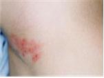
사진 92. 대상포진 피부 발진.
Copyrightⓒ 2001 John Sangwon Lee, MD., FAAP
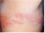
그림93. 대상포진 피부 발진.
Copyrightⓒ 2001 John Sangwon Lee,MD.,FAAP
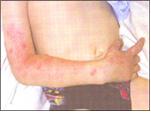
그림94. 6세 된 아이에게 생긴 대상포진
생후 15개월에 수두 예방접종 백신을 받았으나 대상포진이 생겼다.
출처-www.dermatlas.org and APP News. Feb. 2005
-
신체 내 잠재성 수두 바이러스가 피부절과 뇌신경이나 말초신경에 감염되어 생기는 수두 바이러스 신경 피부발진이다.
-
이렇게 생긴 수두 바이러스 신경 피부발진을 수두라고 부르지 않고 대상 포진이라고 한다.
-
신경이 분포된 피부절에 따라 대상포진이 나기 때문에 대상포진은 일정한 피부 부위에 한정돼서 난다.
-
대상포진이 나타나기 전에 그 포진이 날 몸의 부위가 몹시 아플 수 있고 발진이 난 후에도 그 부위가 아플 수 있다.
-
홍반, 구진, 수포 등의 발진이 신체 한쪽의 말초 신경의 분포 주행에 따라 일정한 피부 절에 나는 것이 보통이다.
-
8~12일 정도 지나면 자연히 낫는다.
-
타이레놀 등 진통제로 진통을 시킨다.
-
경구용 Acyclovir로 치료할 수 있다. 소아들의 위장관에서 흡수가 잘 되지 않기 때문에 치료 문제가 생길 수 있다. [부모도 반의사가 되어야 한다–소아가정간호백과]-제7권 소아 청소년 감염병 p000수두와 대상포진 참조.
-
성인들의 대상포진을 Famciclovir 이나 Valacyclovir로 치료할 수 있다.
-
소아들의 대상포진은 선별적으로 항바이러스제로 치료한다.
-
만일 대상포진에 걸린 아이들에게 면역 타협이 있으면 혈관 Acyclovir주사로 치료한다. 출처-www.dermatlas.org and APP News 참조.
11. 단순헤르페스 감염병 (단순포진 감염병) Herpes simplex infection
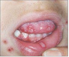
그림95. 단순포진 감염에 의한 치은구강염과 피부염.
Copyrightⓒ 2001 John Sangwon Lee, MD., FAAP
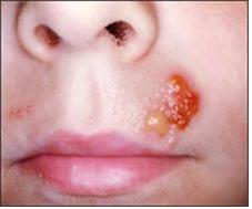
그림96. 단순포진 감염에 의한 피부염.
Copyrightⓒ 2001 John Sangwon Lee, MD., FAAP
-
1형 헤르페스 바이러스 감염으로 인해 홍반, 구진, 수포 등의 발진이 입술, 얼굴, 팔, 다리 몸통, 손가락, 또는 눈 주위의 피부에 생길 수 있다.
-
열이 나지 않거나 미열이 날 수 있다.
-
1형 헤르페스 바이러스 치은구내염을 앓을 때는 고열이 나는 것이 보통이다.
-
1형 단순 헤르페스 치은구내염은 7~10일 정도 되면 자연히 낫는다.
-
이 병이 다 낫을 때쯤 헤르페스 바이러스 피부 발진이 아래 입술 주위에 나타날 수 있다.
-
치은구내염은 없이 헤르페스 단순 피부염만 생길 수 있다
-
2형 헤르페스 감염이 외부 생식 피부도 생길 수 있고 질염도 생길 수 있고, 남성 외부 생식기 헤르페스 피부염 등이 생길 수 있다
-
병력, 증상 징후, 진찰소견 등으로 진단하는 것이 보통이다.
-
대증치료를 하고 경구용 Acyclovir로 치료할 수 있다.
-
[부모도 반의사가 되어야 한다–소아가정간호백과]-제7권 소아 청소년 감염병 p000단순포진 감염(단순 헤르페스 감염)
-
[부모도 반의사가 되어야 한다–소아가정간호백과]-제7권 소아 청소년 감염병-p000 단순헤르페스 치은 구내염 참조.
12. 단핵구증 피부 발진 Mononucleosis skin eruption
-
이비 바이러스나 사이토메갈로바이러스 감염으로 생긴 단핵구증을 앓을 때 피부 발진이 생길 수 있다.
1). 전염성 단핵구증(감염성 단핵구증/전염성 단핵 세포증/모노) Infectious mononucleosis
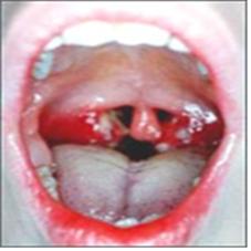
시진 97. 전염성 단핵구증으로 생긴 편도선염.
출처-Copyrightⓒ 2001 John Sangwon Lee, MD., FAAP
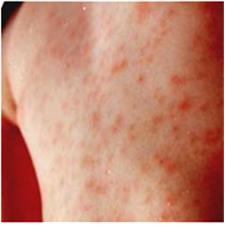
사진 98. 전염성 단핵구증으로 생긴 피부 발진.
출처-Copyrightⓒ 2001 John Sangwon Lee, MD., FAAP
-
이비바이러스 감염으로 생기는 바이러스 감염병을 전염성단핵구증 또는 모노라고 한다.
-
이 병을 앓을 때 열, 인두염, 림프절 비대, 권태감, 상기도염, 간 비대와 비장 비대 등의 증상과 증후가 생기고 구진성 피부 발진이나 다른 형태의 피부 발진이 이병을 있는 환자들의 ⅓에서 생길 수 있다.
-
자주색, 붉은 색, 구리 색 등의 홍반성 구진, 점상 출혈 반점, 두드러기, 다양성 홍반 등이 몸통이나 사지 등에 날 수 있다.
-
이런 발진은 5% 정도 날 수 있고 소양감이 있다.
-
이 병을 앓을 때 Ampicillin이나 Amoxicillin 등 항생제로 치료받으므로 80% 피부 발진이 날 수 있다.
-
병력, 증상 증후, 진찰소견, CBC 피 검사, 모노검사(전염성 단핵구증 검사) 등의 결과로 종합해서 진단하고 대증치료를 한다.
-
[부모도 반의사가 되어야 한다–소아가정간호백과]-제8권 소아청소년 호흡기 질환–전염성 단핵구증 참조.
2). 사이토 메갈로 바이러스 단핵구증 피부 발진 CMV Mononucleosis exanthem
-
사이토메갈로바이러스 감염으로 단핵구증이 생길 수 있다.
-
그 증상 징후는 이비 바이러스 감염으로 생긴 감염성 단핵구증의 증상 징후와 거의 비슷하고 피부 발진도 거의 비슷하다.
-
열이 1~5 주간 계속 날 수 있고
-
Ampicillin이나 Amoxicillin으로 치료하면 80~100% 정도 피부 발진이 생길 수 있다.
-
이 병이 있을 때 모노 검사를 하면 음성으로 나타난다.
-
병력, 증상 징후, 진찰소견을 종합해서 진단하고 대증치료 한다.
Diseases with skin rash and fever(1) 피부 발진과 열이 나는 질병들(1)
• Many children and adolescents (0-18 years old) have a fever and a rash on the skin.
• Here we introduce some of the many ailments that children and adolescents have with fever and rashes on the skin.
• The types, types, and causes of skin rashes, which are relatively common in children and adolescents, have already been explained through skin rash pictures.
• Diseases with only a rash on the skin and no fever
• Fever but no rash on the skin;
• Summarize the skin rash and fever in each of the following sections.
Different types of skin rashes (see photo 2)
• Erythema – red spots caused by congestion in the capillaries
• Papules—a limited, superficial, hard skin rash that rises above the epidermis
• Vesicles – vesicles containing bodily fluid or a rash in which the skin swells up to the size of millet or alveoli, and a skin rash with serous fluid trapped therein.
• Pustules – skin spots formed by the collection of pus within or under the epidermis
• Purpura- Violet or reddish-brown skin spots may or may not be palpable on the skin surface due to inflammation, infection, or bleeding in the walls of blood vessels within the tissue. However, it can be easily seen with the naked eye.
• Petechiae—Slightly raised reddish-purple circular petechiae resulting from intradermal or submucosal hemorrhage.
When children and adolescents (0~18 years of age) only have a skin rash, or if they have a fever and a skin rash, the skin rash may belong to an infectious disease or other disease that falls under the following 1.~29. Rarely, the skin rash may then be caused by one, two, or more than one of several infectious diseases or conditions. Of course, skin rashes can be caused by diseases other than those listed here.
1. Scarlet Fever
2. Meningococcemia and, or Meningococcal meningitis
3. Lyme disease and, or Lyme arthritis
4. Infectious impetigo (Impetigo contagiosa)
5. Toxic Epidermal Necrolysis or Scalded skin syndrome
6. Measles Rubeola (Measles)
7. Rubella (3 days measles)
8. Exanthem subitum/Roseola
9. Chickenpox (Varicella)
10. Erythema infectious/Fifth Disease
11. Herpes Zoster/Shingles
12. Herpes simplex skin infection
13. Infectious mononucleosis skin eruption
14. Hand, foot, mouth disease
15. Adenoviral exanthem
16. Enteroviral exanthem
17. Rotaviral exanthem
18. Pityriasis rosea
19. Erythema multiforme or Steven-Johnson’s syndrome
20. Rocky Mountain spotted fever
21. Asymmetric periflexural exanthem of childhood/Apec
22. Papular-purpuric gloves and socks syndrome
23. Rheumatic fever and, or Rheumatic arthritis
24. Juvenile rheumatoid arthritis
25. Kawasaki disease/Mucocutaneous lymph node syndrome
26. Systemic lupus erythematosus
27. Gianotti-Crosti syndrome
28. There is no fever, but when a certain disease is treated with a drug, a rash and fever may occur on the skin due to the side effects of the drug,
29. In addition, when an infectious disease is treated with a drug, a skin rash may occur as a side effect of the drug and a fever may occur due to the infectious disease.
30. Others
1. Scarlet Fever
• Scarlet fever may occur with pharyngitis or pharyngotonsillitis caused by a group A beta-hemolytic streptococcal infection.
• Rarely, scarlet fever can occur when there is a group A beta-hemolytic streptococcal infection in a skin wound.
• Scarlet fever skin rash is caused by a fungal toxin from a strain of the group A beta-hemolytic streptococci that causes pharyngitis or pharyngotonsillitis.
• You may have a fever, sore throat, aches in your limbs and body, and symptoms such as vomiting and headaches.
• A papule erythematosus rash may appear on the face, neck, chest, stomach, or hands.
• The skin rash can be sandpaper-like. This infectious disease is called scarlet fever.
Figure 76. Group A beta-hemolytic streptococcal tonsillitis and scarlet fever. A red strawberry-shaped tongue is one of the symptoms of scarlet fever. Copyright ⓒ 2011 John Sangwon Lee, MD., FAAP
Figure 75. Red strawberry-shaped tongue due to scarlet fever. Copyright ⓒ 2011 John Sangwon Lee, MD., FAAP
Figure 77. Scarlet fever causes a thin layer of the epidermis of the palm. Copyright ⓒ 2011 John Sangwon Lee, MD., FAAP
Figure 78. When you have scarlet fever, you get a sandpaper-like, reddish skin rash. Health information service, Merk Sharp & Dome, West Point, PA
• The color of the tongue may change to resemble a red strawberry or red beef,
• The pharynx and tonsils are red and the tonsils are enlarged.
• You can easily diagnose this disease by combining your medical history, symptoms, and examination findings.
• Confirmation is confirmed by a group A beta-hemolytic streptococcal antigen antibody agglutination test (Quick Strep Test) or a group A beta-hemolytic streptococcal culture test by collecting mucus from the pharyngeal tonsil. • It should be treated with antibiotics.
•www.drleepediatrics.com- See Volume 7 Infectious Diseases in Children and Adolescents – Scarlet Fever.
2. Lyme disease and Lyme arthritis
Figure 79. Chronic migratory erythema from Lyme disease. Copyright ⓒ 2011 John Sangwon Lee, MD., FAAP
Figure 80. Chronic erythema migratory due to Lyme disease. Copyright ⓒ 2011 John Sangwon Lee, MD., FAAP
• Borrelia Burgdorferi is an infectious disease caused by bacterial infection.
• Arthritis, carditis or meningitis may occur, and chronic Erythema Chronicum Migrans may develop.
• The shape of chronic migratory erythema can be round, oval, etc.
• One or several can fly simultaneously on the face, neck, torso, and legs.
• The central area may be the same as normal skin color.
• The overall color of chronic migratory erythema may be red.
• Chronic erythema migratory also occurs when Lyme disease is present, and the bacteria that cause Lyme disease can directly infect the skin, resulting in primary Lyme disease dermatitis. Source-18
• Diagnosis is based on medical history, symptom signs, examination findings, and Lyme disease test.
• Lyme disease in children and adolescents before the age of 12 is treated with antibiotics such as Amoxicillin, • Lyme disease in children after 12 years of age is treated with antibiotics such as Doxycyclin. • See Lyme disease
3. Meningococcemia and, or Meningococcal meningitis
Figure 81. Skin hemorrhagic spots caused by meningococcal sepsis Used with permission from Ross lab. Columbia Ohio. USA
Figure 82. Purpura and blisters caused by meningococcal sepsis. Source:10
• Meningococcal sepsis is an infectious disease caused by meningococcal infection.
• At first, symptoms are similar to those of a cold, but systemic symptoms such as high fever, headache, low blood pressure, lethargy, and convulsions may develop severely.
• This fungus can directly infect the skin and cause meningococcal dermatitis on the skin. • Erythematous papules and petechiae similar to a measles rash may appear;
• Thrombocytopenia and blood clotting disorders can cause purpura and petechiae throughout the body.
• Diagnosis is made based on medical history, symptom signs, examination findings, and bacterial culture, and treated with appropriate antibiotic vascular injections.
• see meningococcal septic meningitis
• www.drleepediatrics.com – Refer to Volume 7 Infectious Diseases in Children and Adolescents – Sepsis
4. Infectious Impetigo (Infectious Impetigo) Impetigo contagiosa
Photo 83. Impetigo. Copyright ⓒ 2011 John Sangwon Lee, MD., FAAP
• Bacterial dermatitis caused by group A beta-hemolytic streptococcus and/or Staphylococcus aureus infection.
• It occurs mainly on the surface of the skin layer such as the face, neck, extremities, and trunk.
• One or several impetigos can fly at the same time.
• Erythema, blisters, pustules, and crusts (scabs) may develop.
• Bactroban cream, Retapamulin ointment, or Centany (Mupirocin ointment) 2% ointment (Source: Contemporary Pediatrics June 2007) can be applied to the local area with impetigo lesions and treated with oral antibiotics such as Oxacillin. have.
• See impetigo.
5. Staphylococcal scalded skin syndrome (SSSS)
• Burn skin syndrome is an acute skin disease caused by a toxin reaction to Staphylococcus aureus.
• Dermatitis, such as skin with severe second degree burns.
• It is more common in infants and pre-ten-year-old school-age children.
• Lesions such as erythematous spots, epidermis detachment, molting, and dropping may occur on the skin, and systemic symptoms such as malaise, fever, and nervousness may occur simultaneously.
• An erythematous rash like sandpaper may also occur.
• These rashes spread all over the body from the part of the head where hair does not grow, and also occur around the lips and between the fingers.
• If the disease is suspected based on the medical history, symptom signs, and examination findings, a staphylococcal bacterial culture test is performed to diagnose.
• Treat with anti-staphylococcal antibiotics such as Vancomycin, Clindamycin, Oxacillin, and symptomatic treatment. www.drleepediatrics.com – Refer to Volume
8 Children and Adolescent Respiratory Diseases
6. Measles/ Rubeola
Figure 84. Coplic spots on the skin and oral mucosa of measles flowers caused by natural measles. Source-Copyrightⓒ 2001 John Sangwon Lee, MD.FAAP
• It is a systemic viral infection caused by measles virus infection.
• The typical symptoms of measles are fever and malaise for the first 24-48 hours after onset, followed by fever and malaise.
o Cough,
o runny nose (Coryza),
o Three symptoms of conjunctivitis (3CS) are always present.
• These three symptoms are called 3Cs symptoms in English.
• Koplik spots on the mucous membranes of the mouth.
• When the spots appear on the 1st or 2nd day, an erythematous papular rash (measles flower) starts from the skin behind the pinna and spreads to the face, trunk, limbs and hands for 2-4 days.
• The skin rash usually goes away in the order in which it appeared over a period of 5 to 8 days.
• Mild measles in children who have been vaccinated against the measles vaccine and have little or no antibodies after they have developed measles immunity, or natural measles and measles in children who have not received the measles vaccine Since the flowers are different, it is necessary to make a differential diagnosis.
• See Measles.
• Diagnosis is made by combining medical history, symptoms, and examination findings. treat symptomatically.
7. Rubella (3 days measles)
• A systemic acute viral infection caused by rubella virus infection is called rubella. 1. headache,
2. My eyes hurt
3. sore throat,
4. Low fever,
5. Minor symptoms such as KEPCO continue for 3 to 4 days.
6. The lymph nodes in the neck area under the occipital bone, behind the pinna and in the neck area below it may become enlarged and painful.
7. Within 4 to 5 days after the onset of this disease, a pale pink rash, erythematous rash and papules start appearing on the forehead and behind the ears, on the areas where hair does not appear on the wheels, and spread to the skin of the face, neck, trunk, stomach, limbs, etc. It spreads.
8. These spots disappear naturally after 2-3 days.
• Diagnosis is made by synthesizing medical history, symptoms, symptoms, and examination findings.
• If the disease occurs during the first trimester of pregnancy, the fetus can develop congenital rubella syndrome.
• The fetus may develop congenital rubella syndrome such as cataracts, congenital heart malformations, deafness, pneumonia, enlarged liver and spleen, and thrombocytopenia.
• www.drleepediatrics.com – Volume 6 Diseases with Good Growth and Development in Newborns – Congenital Rubella Syndrome. See Volume 7, Infectious Diseases in Children and Adolescents – Rubella
Figure 85. A skin rash caused by rubella and enlarged lymph nodes behind the pinna and at the edge of the scalp. source; Health information service, Merk Sharp & Dome, West Point, PA
Picture 86. Enlarged lymph nodes behind the auricle and at the edge of the scalp due to rubella. Copyright ⓒ 2011 John Sangwon Lee, MD. FAAP
8. Exanthem subitum (Roseola)
Acute viral infections caused by human herpesvirus 6 infection or human herpesvirus 7 infection.
• It is an infectious disease that mainly affects children aged 3 months to 3 years. • A high fever of 39 to 40 degrees Celsius continues for 1 to 4 days, and as soon as the fever subsides, red erythematous spots similar to measles flowers or rubella flowers, erythematous papules may appear.
• It is common for this rash to appear first on the torso and then spread to the face, neck, and limbs.
Fig. 87. Skin rash on the rosette. Copyright© 2001 John Sangwon Lee, MD.FAAP
• The rash progresses for 1 to 2 days and then goes away on its own.
• A high fever can cause febrile convulsions before the rash occurs.
• It is often difficult to confirm the diagnosis because there are no specific symptoms on examination.
• From the day of the onset to the time the fever subsides, it can be quite refreshing.
• Diagnosis is made by synthesizing medical history, symptom signs, and examination findings, and fever is treated with Tylenol.
•www.drleepediatrics.com – See Volume 7 Infectious Diseases in Children and Adolescents – Sudden Rash.
9. Erythema Infectiosum (Fifth Disease/Human parvovirus B19 infection and Skin rashes)
• It is a viral infection caused by human parvovirus B19 infection.
• This disease is also called the fifth disease.
• Symptoms such as fever, headache, cold sores, muscle pain, and malaise may occur slightly.
• At first, both cheeks are red, and then erythematous or papule-like spots may appear on both shoulders and upper arms, similar to a net or lace-like appearance.
• The rash may go on for two to three weeks.
• About 10% of joint pain occurs.
• If a pregnant woman becomes infected with this virus, the fetus may develop severe anemia or hydrocephalus.
• Diagnosis is made by synthesizing medical history, symptoms, signs, and examination findings.
• Treat symptomatically
• Parents should also become anti-doctors-Children’s Family Encyclopedia]-See Volume 7 Infectious Diseases in Children and Adolescents.
10. Chickenpox Chickenpox (Varicella)
Figure 91. Chickenpox skin rash. Copyright© 2001 John Sangwon Lee, MD.FAAP
Figure 90. Chickenpox skin rash. Copyright© 2001 John Sangwon Lee, MD.FAAP
• It is a viral disease caused by varicella virus infection.
• The disease develops after an incubation period of 10 to 20 days.
• Systemic symptoms such as mild to high fever and headache may occur.
• At first, an erythematous rash appears on the body, then the rash turns into a papule-like rash, and then the chickenpox rash progresses in the order of blisters, pustules, and crusts.
• Various types of chickenpox rash can be seen at once, and the phenomenon that it starts on the body first and spreads to the limbs and face is a peculiarity of chickenpox rash.
• After 5 to 10 days, a rash appears, and the rash heals as it scabs.
• When a skin rash occurs, it is usually accompanied by symptoms such as itching and malaise.
• After the chickenpox rash has cleared up, there may be small scars. • natural chickenpox due to infection with the natural chickenpox virus;
• Vaccination varicella due to live varicella virus infection in the varicella vaccine;
• After receiving the chickenpox vaccine, the chickenpox immune system is protected against chickenpox for a while and then the antibody concentration naturally decreases. Natural chickenpox caused by infection with the natural chickenpox virus should be differentially diagnosed.
• Symptoms and signs of this type of chickenpox are different.
• Diagnosis is made by combining medical history, symptoms, and examination findings.
• Antipyretic, analgesic and anti-inflammatory with Tylenol.
• Treat with Acyclovir (Zovirax) as needed
• www.drleepediatrics.com – See Volume 7 Infectious Diseases in Children and Adolescents – Chickenpox and Herpes Zoster 11. Herpes Zoster (Shingles)
Picture 92. Herpes zoster skin rash. Copyright© 2001 John Sangwon Lee, MD., FAAP
Figure 93. Herpes zoster skin rash. Copyrightⓒ 2001 John Sangwon Lee, MD.,
FAAP
Figure 94. Shingles in a 6-year-old child She received the chickenpox vaccine at 15 months of age, but developed shingles. Source – www.dermatlas.org and APP News. Feb. 2005
• It is a varicella virus nerve skin eruption caused by an infection of the body’s latent varicella virus in the dermatome, cranial or peripheral nerves.
• The chickenpox virus nerve skin rash that looks like this isn’t called chickenpox, it’s called herpes zoster.
• Because herpes zoster occurs according to the innervated dermatome, herpes zoster is limited to a certain area of skin.
The area of the body where the shingles will strike can be very painful before the onset of shingles, and the area can be sore after the rash.
• It is common for rashes such as erythema, papules, and blisters to appear in certain skin nodes according to the distribution of peripheral nerves on one side of the body.
• It will heal naturally after 8 to 12 days.
• Pain relief with pain relievers such as Tylenol. • It can be treated with oral Acyclovir.
Treatment problems can arise because of poor absorption from the gastrointestinal tract in children.
www.drleepediatrics.com – See Volume 7 Infectious Diseases in Children and Adolescents p000 Chickenpox and Herpes Zoster.
• Herpes zoster in adults can be treated with Famciclovir or Valacyclovir. • Herpes zoster in children is selectively treated with antiviral drugs.
• If there is compromised immunity in children with shingles, treat with vascular Acyclovir. Source – see www.dermatlas.org and APP News.
11. Herpes simplex infection
Figure 95. Gingivitis and dermatitis caused by herpes simplex infection. Copyright© 2001 John Sangwon Lee, MD., FAAP
Figure 96. Dermatitis caused by herpes simplex infection. Copyright© 2001 John Sangwon Lee, MD., FAAP
• Herpes 1 virus infection can cause rashes such as erythema, papules, and blisters on the lips, face, arms, trunk of legs, fingers, or the skin around the eyes. • You may not have a fever or have a slight fever.
• It is normal to have a high fever when you have herpes type 1 virus gingivitis.
• Type 1 herpes simplex gingivostomatitis heals spontaneously in 7 to 10 days.
• By the time the disease is cured, a herpes virus skin rash may appear around the lower lip.
• Only herpes simplex dermatitis can occur without gingivostomatitis.
• Herpes type 2 infection can also cause external genital skin, vaginitis, and male external genital herpes dermatitis.
• Diagnosis is usually based on medical history, symptoms, and examination findings.
• It can be treated symptomatically and with oral Acyclovir.
• [Parents should also become anti-doctors – Encyclopedia of Pediatric and Family Nursing] – Vol. 7 Infectious disease herpes simplex infection (herpes simplex infection) in children and adolescents
• www.drleepediatrics.com-Volume 7 Infectious diseases in children and adolescents-p000 See herpes simplex gingival stomatitis.
12. Mononucleosis skin eruption
• A skin rash may occur if you have mononucleosis caused by an ovary virus or cytomegalovirus infection.
1). Infectious mononucleosis (infectious mononucleosis/infectious mononucleosis/mono) Examination
97. Tonsillitis caused by infectious mononucleosis. Source-Copyrightⓒ 2001 John Sangwon Lee, MD., FAAP
Picture 98. Skin rash caused by infectious mononucleosis. Source-Copyrightⓒ 2001 John Sangwon Lee, MD., FAAP
• Viral infectious disease caused by ENT virus infection is called infectious mononucleosis or mono.
• Symptoms and symptoms such as fever, pharyngitis, enlarged lymph nodes, malaise, upper respiratory tract infection, hepatomegaly and spleen enlargement, and papular skin rash or other skin rashes may occur in one-third of patients with this disease. have.
• Purple, red, copper-colored erythematous papules, petechiae spots, urticaria, and erythema multiforme may appear on the trunk or extremities.
• These rashes can be 5% of the time and are itchy.
• When you have this disease, you are treated with antibiotics such as Ampicillin or Amoxicillin, so 80% of skin rashes can occur.
• Comprehensive diagnosis and symptomatic treatment based on the results of medical history, symptom symptoms, examination findings, CBC blood test, and monotest (infectious mononucleosis test).
• www.drleepediatrics.com – See Volume 8 Children and Adolescent Respiratory Diseases – Infectious Mononucleosis.
2). Cytomegalovirus mononucleosis skin rash CMV Mononucleosis exanthem
• Cytomegalovirus infection can cause mononucleosis.
• The symptoms are very similar to those of infectious mononucleosis caused by an ENT virus infection, and the skin rash is almost identical.
• Fever can last for 1 to 5 weeks
• Treatment with Ampicillin or Amoxicillin may cause skin rashes in 80-100% of cases.
• When you have this disease, the mono test is negative.
• Diagnose and treat symptomatically by combining medical history, symptoms, and examination findings.
출처 및 참조문헌
- www.drleepediatrics.com제6권 신생아 성장 발육 육아 질병
- www.drleepediatrics.com제7권 소아청소년 감염병
- www.drleepediatrics.com제8권 소아청소년 호흡기 질환
- www.drleepediatrics.com제9권 소아청소년 소화기 질환
- www.drleepediatrics.com제10권. 소아청소년 신장 비뇨 생식기 질환
- www.drleepediatrics.com제11권. 소아청소년 심장 혈관계 질환
- www.drleepediatrics.com제12권. 소아청소년 신경 정신 질환, 행동 수면 문제
- www.drleepediatrics.com제13권. 소아청소년 혈액, 림프, 종양 질환
- www.drleepediatrics.com제14권. 소아청소년 내분비, 유전, 염색체, 대사, 희귀병
- www.drleepediatrics.com제15권. 소아청소년 알레르기, 자가 면역질환
- www.drleepediatrics.com제17권. 소아청소년 피부 질환
- Red book 29th-31st edition 2021
- Nelson Text Book of Pediatrics 19th — 21st Edition
- The Johns Hopkins Hospital, The Harriet Lane Handbook, 22nd edition
-
Childhood Emergencies in the Office, Hospital and Community, American Academy of Pediatrics
-
Emergency Medical Service for Children, By Ross Lab. May 1989. p.10
-
Emergency care, harvey grant and robert murray
-
Emergency Care Transportation of Sick and Injured American Academy of Orthopaedic Surgeons
-
Emergency Pediatrics A Guide to Ambulatory Care, Roger M. Barkin, Peter Rosen
-
Immediate care of the acutely ill and injured, Hugh E. Stephenson, Jr
-
The Critically Ill Child, Diagnosis and Management, Edited by Clement A. Smith
-
Emergency Medical Services for Children: The Role of the Primary Care Provider, America Academy of Pediatrics
-
Quick Reference To Pediatric Emergencies , Delmer J. Pascoe, M.D., Moses Grossman, M.D. with 26 contributors
-
Manual of Emergency Care
-
응급환자관리 정담미디어
-
소아가정간호백과–부모도 반의사가 되어야 한다, 이상원
-
Neonatal Resuscitation American heart Association
-
Neonatology Jeffrey J.Pomerance, C. Joan Richardson
-
Pediatric Resuscitation Pediatric Clinics of North America, Stephen M. Schexnayder, M.D.
-
Pediatric Critical Care, Pediatric Clinics of North America, James P. Orlowski, M.D.
-
Preparation for Birth. Beverly Savage and Dianna Smith
- Infectious disease of children, Saul Krugman, Samuel L Katz, Ann A. Gershon, Catherine Wilfert
-
The Harriet Lane Handbook 19th Edition
-
소아과학 대한교과서
-
제1권 소아청소년 응급의료 참조문헌과 출처
-
Other
다음은 피부 발진과 열이 나는 질병들(2) Diseases with skin rash and fever(2)로 이어짐
Copyright ⓒ 2015 John Sangwon Lee, MD., FAAP
“부모도 반의사가 되어야 한다”-내용은 여러분들의 의사로부터 얻은 정보와 진료를 대신할 수 없습니다.
“The information contained in this publication should not be used as a substitute for the medical care and advice of your doctor. There may be variations in treatment that your doctor may recommend based on individual facts and circumstances. “Parental education is the best medicine.”