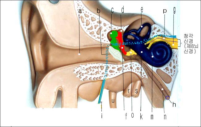
정상 외이, 중이와 내이 그림 Diagram of a normal outer, middle, and inner ear
정상 외이, 중이와 내이 그림
Diagram of a normal outer, middle, and inner ear
a- 외이도, b-고막, c-추골, d-중이(중이강), e-반규관(반고리관), f-침골, g-와우신경, h-이관 입구, i-안면신경(제 7뇌신경), k-전정, m-와우, n-이관(구씨관), o-등골, p-전정신경(전정와우신경/제 8뇌신경), r-경정맥, s-내경동맥, u-경돌상돌기
Diagram of a normal outer, middle, and inner ear
a- External auditory canal, b- Tympanic membrane, c- Malleus, d- Middle ear (tympanic cavity), e- Semicircular canals, f- Incus, g- Cochlear nerve, h- Opening of the Eustachian tube, i- Facial nerve (7th cranial nerve), k- Vestibule, m- Cochlea, n- Eustachian tube, o- Stapes, p- Vestibular nerve (vestibulocochlear nerve/8th cranial nerve), r- Jugular vein, s- Internal carotid artery, u- Mastoid process
Copyright drleepediatrics.com 1/29/2026
Copyright drleepediatrics.com 3/8/2026