딸기 혈관종(모세 혈관종/딸기양 혈관종)
Strawberry hemangioma(Capillary hemangioma/Strawberry nevus)
For more information, please visit drleepediatrics.com. search 정보를 더 찾으려면 drleepediatrics.com 방문한후 search 에서 찾아보세요.
● 딸기 혈관종은 빨간 딸기색과 같이 혈관종이 빨갛고, 피부의 표면 위로 현저히 솟아 나 있고, 혈관종의 경계가 뚜렷한 혈관성 모반의 일종이다.
▴ 사진485. 딸기 혈관종.
Copyrightⓒ 2001 John Sangwon Lee, MD.FAAP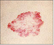
▴ 사진 486. 딸기 혈관종이 자연히 더 나아가는 과정.
Copyrightⓒ 2001 John Sangwon Lee, MD.FAAP
▴ 사진 488. 딸기 혈관종이 자연히 거의 다 나아가는 과정.
Copyrightⓒ 2001 John Sangwon Lee, MD.FAAP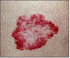
▴ 사진 487. 딸기 혈관종이 자연히 나아가는 과정.
Copyrightⓒ 2001 John Sangwon Lee, MD.FAAP
● 혈관성 모반 중 가장 흔한 종류이다.
● 딸기 혈관종을 딸기양 모반, 딸기 혈관종, 또는 모세 혈관종이라고도 한다(사진 485~488 참조).딸기 혈관종은 출생 후 바로 육안으로 볼 수 있을 정도로 확연히 나타나기도 한다.
● 그렇지만 대부분의 경우 생후 1~2개월 경 더 현저히 나타나기 시작한다.
● 그 후부터는 점점 더 현저히 나타나고, 또 점점 더 커지다가 생후 1~2세경부터 점점 작아지기 시작한다.
● 그 후 계속 점점 더 작아지다가 7~8세경에는 자연히 완전히 없어지는 것이 자연 진행 과정이다.
● 딸기 혈관종에서 이런 자연 진행 과정을 따르는 빈도는 70~80% 정도이다.
● 전 딸기 혈관종의 70~80%는 7~8세경에 자연히 없어진다.
● 신체의 어느 부위의 피부에도 생길 수 있지만 얼굴, 머리, 가슴, 사지 등의 피부에 더 잘 난다.
■ 딸기 혈관종(모세 혈관종/딸기양 혈관종) 증상 증후
● 대부분의 딸기 혈관종은 아무 증상이 없고 보기에 이상할 뿐이다.
● 그렇지만 눈꺼풀이나 콧구멍 근처, 또는 귓구멍 근처에 크게 나서 그 부위에 있는 귓구멍, 콧구멍을 막아 그로 인해 그 기관의 기능에 장애가 생길 수 있다.
● 안구 주위에 난 딸기 혈관종으로 안구가 가려지면 그 가려진 정도에 따라 약시 등 시야장애가 생길 수 있다.
● 기저귀를 채우는 부위에 난 딸기 혈관종에 궤양이 생기기 쉽다.
● 궤양이 생긴 혈관종에 세균 감염이 생길 수 있고 또 혈관종 궤양에서 출혈할 수 있다.
● 턱수염이 나는 부위에 딸기 혈관종이 나 있으면 기도 속에도 혈관종이 나 있을 확률이 63% 정도나 된다.
● 코 끝 부분에 난 딸기 혈관종이나 입술에 난 딸기 혈관종은 미관상 문제가 될 수 있다.
● 손에 난 딸기 혈관종을 손 기능 장애가 생길 수 있다.
● 딸기 혈관종이 신생아에게 많이 나 있을 때는 그들의 간, 폐, 뇌, 소화기 등, 내부 장기에도 혈관종이 나 있을 가능성이 있다
● 일반적으로 딸기양 모반은 신생아기 동안에는 아주 작지만 생후 1~2년 간 점점 더 커지고 색깔도 점점 더 빨갛게 변하는 것이 보통이다.
● 그렇지만 생후 1~2년 이후부터 딸기 혈관종이 점점 더 작아지기 시작해서 3세에 30%, 5세에 50%, 9세에 90% 이상이 자연히 사라지는 것이 보통이다.
■ 딸기 혈관종(모세 혈관종/딸기양 혈관종) 치료
● 대부분의 딸기 혈관종은 생후 1~2년이 지난 후부터 생후 7세 까지 몇 년간을 거쳐 점점 작아지면서 자연히 사라진다.
● 따라서 대개의 경우 두고 관찰적 치료를 하는 것이 보통이다.
● 귓구멍이나 콧구멍의 주위에 나서 귓구멍이나 콧구멍을 막아 그 기관의 기능 장애를 일으키는 딸기 혈관종이나, 눈꺼풀이나 눈 주위에 나서 안구를 가리는 딸기 혈관종, 호흡기에 난 딸기 혈관종, 궤양성 혈관종 등 기능장애를 일으키는 딸기 혈관종은 의사의 처방에 따라 전신성 경구용 코르티코스테로이드제로 몇 개월 동안 치료하기도하고 전신성 펄스드 혈관 코르티코스테로이드제 치료를 할 수 있다.
● 전신 경구용 코르티코스테로이드제로 치료한 결과가 전신 혈관 코르티코스테로이드제로 치료한 결과 보다 더 낫지만 부작용이 더 많다고 한다30.
● 전신성 경구용 코르티코스테로이드제 치료의 효과는 8% 정도 된다고 한다.
● 그 외 레이저 치료, 또는 Interferon-알파 치료도 한다.
● 딸기 혈관종의 5~15%에서 혈관종에 궤양이 생길 수 있다.
● 궤양의 정도에 따라 항생제 연고나 크림으로 치료하거나, 외과적 수술치료, 펄스드 다이 레이저 치료, 국소 코르티 코르티코스테로이드제 주사와 전신 코르티코스테로이드제로 치료하기도 한다22.
● 그 밖의 신체 다른 피부에 생긴 것은 나이에 따라 점점 더 커지다가 자연히 없어지므로 그대로 놓고 관찰한다.
■ 혈관종의 합병증
Complications of Hemangioma
● 대부분의 혈관종은 합병이 생기지 않으나 때로는 다음과 같은 합병증이 생길 수 있다.
● 혈관종에 궤양이 생길 수 있다.
● 가장 흔히 볼 수 있는 합병증이다.
● 궤양이 생기면 아플 수 있고 거기에 박테리아 감염이 생길 수 있고 출혈도 생길 수 있다. 궤양이 나은 후 상흔이 생길 수 있다.
● 봉소염, 골수염, 또는 패혈증도 생길 수 있다.
● 혈관종이 외이도 입구에 생기면 중이염도 생길 수 있고 소리 전도 장애 난청이 생길 수 있다.
● 안구 주위에 혈관종이 생겨 안구와 시야가 막히면 약시 또는 난시가 생길 수 있다.
● 혈관종이 안면에 여러 개 나 있을 때나 상당히 큰 혈관종이 안면에 나 있으면 혈관종이 간이나 호흡기계, 또는 비뇨생식기계에도 혈관종이 나 있을 수 있다.
● 큰 혈관종이 안면에 나 있을 때 두개 강내 혈관종이 나 있을 수 있다.
● 턱, 입술, 하악, 목 등에 혈관종이 나 있으면 성문에도 혈관종이 나 있을 수 있다.
● 요천추 부위의 피부에 혈관종이 나 있으면 척수, 항문직장, 비뇨생식기계에도 혈관종이 나 있을 수 있다.
● 카사바하-메리트 증후군도 생길 수 있다.
● 그 외
■ 혈관종의 진단 치료
병력, 증상 징후, 검진, 피검사, 울트라사운드 검사로 진단한다.
Vincristine, Propranolol,또는 Regranex (becaplermin) gel 등으로 치료도 한다.
● Hemangeol (propranol hydrochloride) 경구용 용액으로 치료하면 3개월 내 80% 완전 치료 된다 9/1/2019 AAP News.
■ 출처 및 참조문헌
● The Johns Hopkins Hospital, The Harriet Lane Handbook, 18th edition, pp 226
● Clinical Pediatric Dermatology, A Textbook of Skin Disorders of Childhood and Adolescence, Sidney Hurwitz, Saunders
Copyright drleepediatrics.com 2/21/2026
Strawberry hemangioma (Capillary hemangioma/Strawberry nevus)
For more information, please visit drleepediatrics.com. Search
For more information, visit drleepediatrics.com and search.
● A strawberry hemangioma is a type of vascular nevus characterized by a red, strawberry-colored hemangioma, prominently raised above the skin surface, and distinct borders.
▴ Photo 485. Strawberry hemangioma.
Copyrightⓒ 2001 John Sangwon Lee, MD.FAAP
▴ Photo 486. Strawberry hemangioma progressing naturally.
Copyrightⓒ 2001 John Sangwon Lee, MD.FAAP
▴ Photo 488. Strawberry hemangioma progressing naturally.
Copyrightⓒ 2001 John Sangwon Lee, MD.FAAP
▴ Photo 487. Natural progression of a strawberry hemangioma.
Copyrightⓒ 2001 John Sangwon Lee, MD.FAAP
● This is the most common type of vascular nevus.
● Strawberry hemangioma is also called strawberry nevus, strawberry hemangioma, or capillary hemangioma (see Photos 485-488). Strawberry hemangiomas can be clearly visible immediately after birth.
● However, in most cases, they become more noticeable around 1-2 months after birth.
● From then on, they become increasingly prominent and larger, before gradually shrinking around 1-2 years of age.
● They then continue to shrink until they disappear completely by 7-8 years of age, a natural progression.
● This natural progression is the frequency of strawberry hemangiomas. ● 70-80% of strawberry hemangiomas disappear spontaneously by age 7-8.
● They can develop on any part of the body, but are more common on the face, head, chest, and extremities.
■ Strawberry Hemangiomas (Capillary Hemangiomas/Strawberry Hemangiomas) Symptoms and Signs
● Most strawberry hemangiomas are asymptomatic and simply appear odd.
● However, if they grow large near the eyelids, nostrils, or ear canals, they can block the ear or nostrils, potentially disrupting the function of those organs.
● If a strawberry hemangioma around the eyeball obscures the eyeball, it can cause visual impairment, such as amblyopia, depending on the degree of obstruction.
● Strawberry hemangiomas in the diaper area are prone to ulceration.
● Ulcerated hemangiomas can become infected, and the hemangioma ulcers can bleed. ● If a strawberry hemangioma is present in the beard area, there’s a 63% chance that another hemangioma is present in the airway.
● Strawberry hemangiomas on the tip of the nose or lips can be cosmetically problematic.
● Strawberry hemangiomas on the hands can cause hand dysfunction.
● When a newborn has a high incidence of strawberry hemangiomas, there’s a chance they’ll also be present in internal organs, such as the liver, lungs, brain, and digestive system.
● Strawberry nevi are typically very small during the newborn period, but they typically grow larger and turn redder over the first year or two of life.
● However, strawberry hemangiomas typically begin to shrink after one or two years of age, with 30% of cases disappearing by age three, 50% by age five, and over 90% by age nine. ■ Treatment for Strawberry Hemangiomas (Capillary Hemangiomas/Strawberry Hemangiomas)
● Most strawberry hemangiomas gradually shrink and disappear naturally over several years, from 1-2 years of age to around 7 years of age.
● Therefore, observational treatment is usually recommended in most cases.
● Strawberry hemangiomas that cause functional impairment, such as those that occur around the ear or nostrils and block their function, those that occur around the eyelids or eyes and obscure the eyeball, those that occur in the respiratory tract, and those that occur as ulcerative hemangiomas, can be treated with systemic oral corticosteroids for several months or with pulsed systemic corticosteroids, depending on the doctor’s prescription.
● While treatment with systemic oral corticosteroids is more effective than treatment with systemic vascular corticosteroids, it is associated with more side effects.30
● Systemic oral corticosteroids are said to be effective in approximately 8% of cases. ● Other treatments include laser therapy or interferon-alpha therapy.
● Strawberry hemangiomas can ulcerate in 5-15% of cases.
● Depending on the severity of the ulcer, treatment may include antibiotic ointment or cream, surgical treatment, pulsed dye laser therapy, topical corticosteroid injections, and systemic corticosteroids.22
● Other skin tumors on the body tend to grow larger with age and resolve naturally, so they should be left alone and observed.
■ Complications of Hemangioma
● Most hemangiomas do not cause complications, but the following complications can sometimes occur:
● Hemangiomas can ulcerate.
● This is the most common complication.
● Ulcers can be painful, can become infected with bacteria, and can bleed. Scarring can occur after the ulcer heals.
● Cellulitis, osteomyelitis, or sepsis can also occur. ● If a hemangioma develops at the entrance to the external auditory canal, it can cause otitis media and hearing loss due to sound conduction disturbances.
● If a hemangioma develops around the eye and blocks the eye and visual field, it can cause amblyopia or astigmatism.
● If multiple hemangiomas are present on the face, or if a relatively large hemangioma is present, hemangiomas may also be present in the liver, respiratory tract, or genitourinary tract.
● If a large hemangioma is present on the face, an intracranial hemangioma may also be present.
● If a hemangioma is present on the chin, lips, mandible, or neck, the glottis may also be present.
● If a hemangioma is present on the skin of the lumbosacral region, it may also be present in the spinal cord, anorectum, or genitourinary tract.
● Casabha-Merritt syndrome may also develop.
● Other
■ Diagnosis and Treatment of Hemangiomas
Diagnosis is based on medical history, symptoms, signs, physical examination, blood tests, and ultrasound examination. Treatment with vincristine, propranolol, or Regranex (becaplermin) gel is also recommended.
● Treatment with Hemangeol (propranol hydrochloride) oral solution results in 80% complete cure within 3 months. 9/1/2019 AAP News.
■ Sources and References
● The Johns Hopkins Hospital, The Harriet Lane Handbook, 18th edition, pp. 226
● Clinical Pediatric Dermatology, A Textbook of Skin Disorders of Childhood and Adolescence, Sidney Hurwitz, Saunders
Copyright drleepediatrics.com 2/21/2026